Get clearer answers with accurate, replicable Western blots and many other assays.
Get a Quote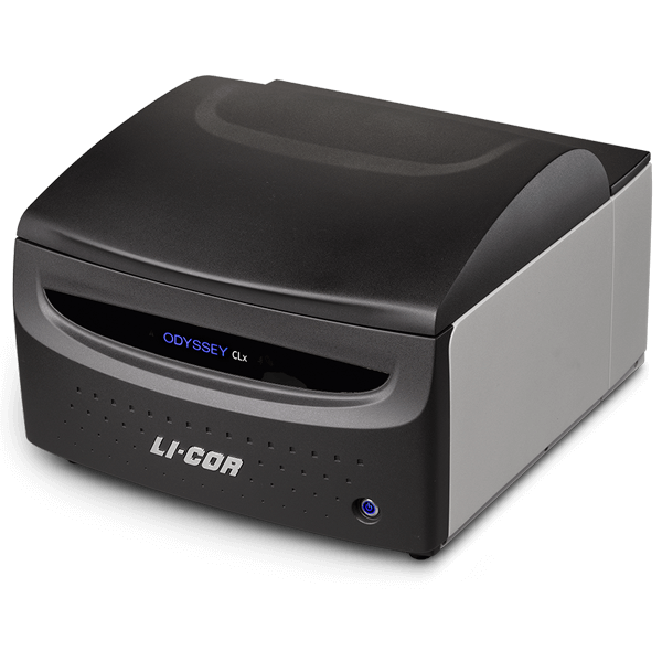
LI-COR considers it a great privilege to provide the products, tools, and services researchers need to arm themselves for the fight against SARS-CoV-2 and other viruses.
The Odyssey CLx Viral Solution Package is currently available for priority delivery and at a reduced price. Installation and training can be done virtually with a Solutions & Support Scientist.
Contact us

Contact us for additional information about the Odyssey CLx Viral Solution package.
*Offer may not be available in all regions.
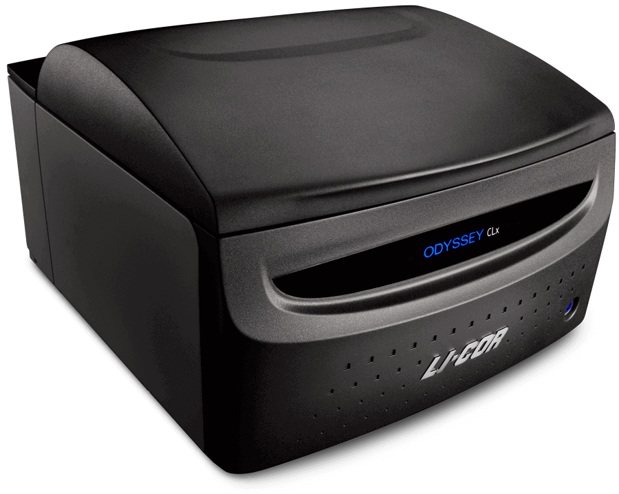
Get consistent, accurate digital images, without the hassles and unpredictability of film – so you can analyze your data right away and plan your next experiment.
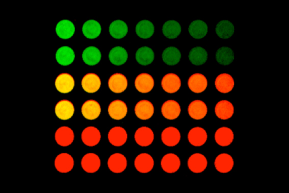
In-Cell Western™ Assay
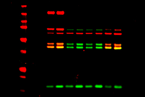
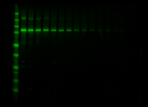
NIR Fluorescent Western Blot
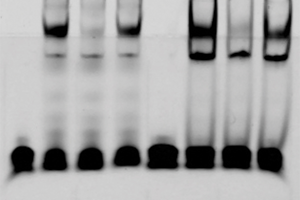
EMSA / Gel Shift Assay
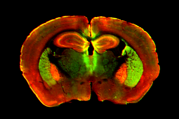
Tissue Section
Image courtesy of C. Kearn,
University of Washington
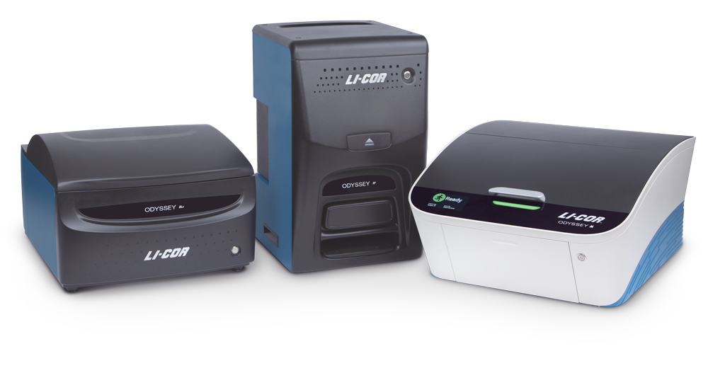
Western blots, cell-based assays, protein arrays, gel shift assays, tissue section imaging, and more are at your fingertips with near-infrared (NIR) fluorescence on the Odyssey CLx.
“We are finding that we can use the Odyssey CLx for just about everything protein-related we do in the lab.”
Analyze your data right away – it’s already in digital format. One digital file contains all your image data. Record-keeping is easy and multiple exposures are a thing of the past.
To image and analyze many samples efficiently, use the Odyssey CLx to scan up to 9 mini-blots, 6 microplates, or 30 slides at the same time. The large scanning bed provides high throughput for many assays.
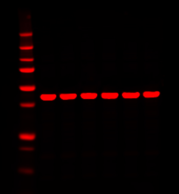
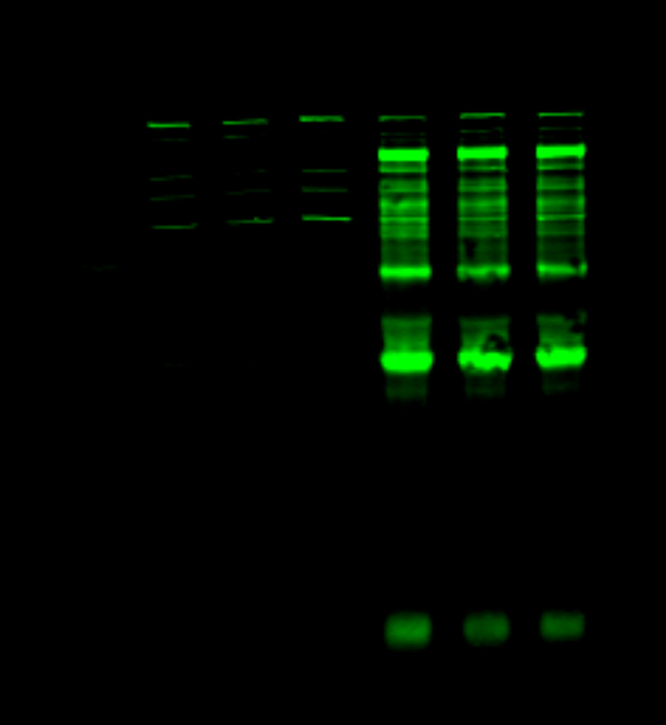
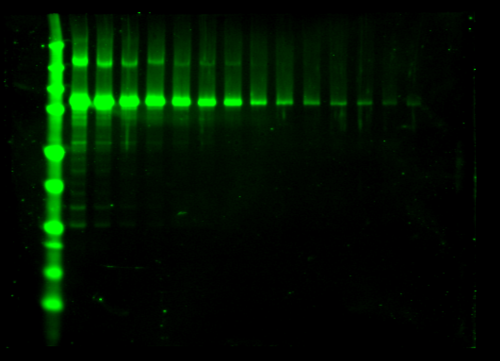
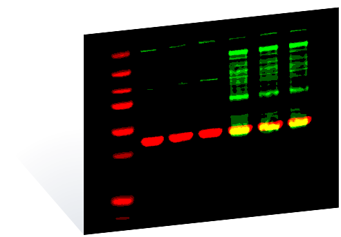
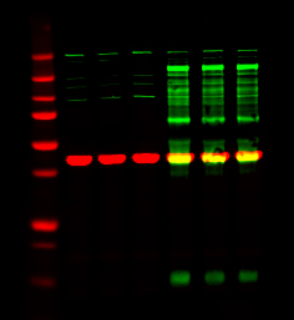
See both strong and faint bands clearly in a single image acquired on the Odyssey CLx, with great sensitivity and no image saturation. Multiplex with two fluorescent colors to see even more data.
One digital image file contains all your data so you can see both strong and faint bands clearly in a single image, without image saturation, “blowout”, or sacrificing sensitivity. Increase the consistency and reliability of your results by capturing a single image with the Odyssey CLx, rather than comparing multiple exposures captured under variable conditions.
“The superior dynamic range and sensitivity allows me to confidently report my data.”


One image file contains the full range of your data, and changing image display settings never alters your raw data or signal intensities.
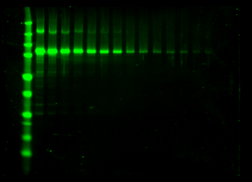
Powerful, precise laser excitation and specialized optics enable the Odyssey CLx to provide high signal-to-noise ratios and outstanding image quality.
High signal-to-noise ratios give you more confidence in your data and can help you reliably detect subtle changes between samples. Low background fluorescence at NIR wavelengths means high sensitivity for your blots.
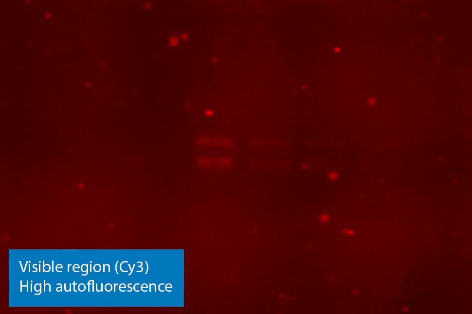
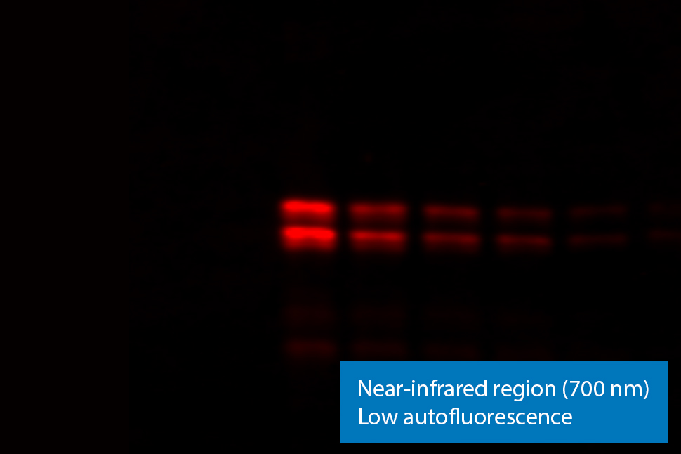
NIR fluorescence imaging gives you low membrane fluorescence and high sensitivity. Other imagers use LEDs or diffuse white light sources that provide weak, non-specific excitation light and limit sensitivity. Visible fluorescent dyes like Cy3 have poor sensitivity, because membrane autofluorescence is so strong. Blots show detection of phospho-ERK with NIR fluorescence (700 nm) and with ECL Plex reagents.
“The brilliant signal-to-noise ratio in combination with the ability to truly quantify the data is really outstanding.”
Enhance the accuracy of your data. The wide dynamic range of Odyssey imagers enables high-sensitivity detection without instrument saturation.
Fluorescent signals are stable and unaffected by timing, so you can compare band intensities with confidence. You can even conveniently re-image the same blot on the Odyssey CLx later and see the same results.
NIR fluorescent signals are stable for months, because no enzymes or substrates are used. Direct detection is performed using secondary antibodies labeled with near-infrared fluorescent dyes, such as IRDye® secondary antibodies. You’ll typically lose signal in less than an hour with ECL substrates and indirect enzymatic detection.
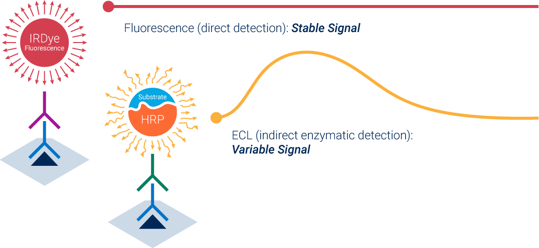
Compare band intensities confidently with direct detection of stable fluorescent signals.
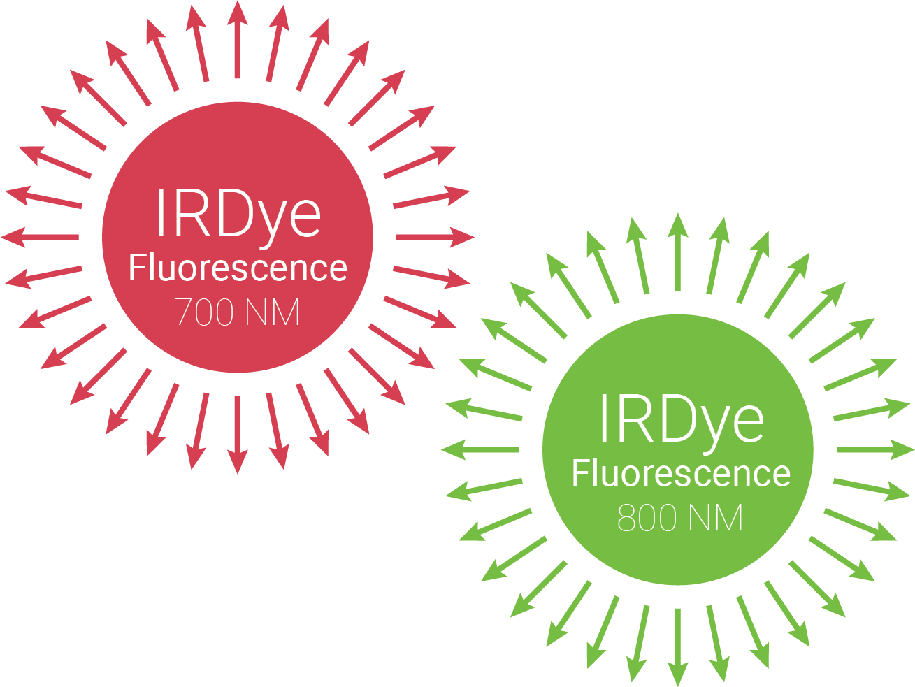
With multiplex fluorescence, you can detect two protein targets in each sample lane, with great sensitivity in both fluorescence channels.
Use secondary antibodies labeled with spectrally-distinct NIR fluorescent dyes to get more data from your blot. View, adjust, and analyze your results as a merged image, or as separate 700 nm and 800 nm channel images in pseudo-color or grayscale. Consistency and accuracy are improved because you can account for lane-to-lane variation in loading and transfer without stripping and re-probing.
“The infrared imaging system allows detection of multiple proteins on a single blot, without the need for stripping and re-probing.”
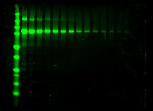
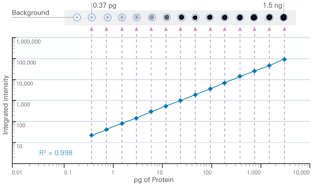
Get deeper data capacity with digital fluorescence and the unprecedented linear dynamic range of the Odyssey CLx, and never lose data because of image saturation.
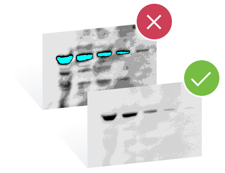
Never lose your data because of image saturation. Digital fluorescent detection with the Odyssey CLx shows your strong signals as they really are. Film and CCD imagers only show you part of the picture, because data are lost when images become saturated. To see the whole picture, you need enough capacity (dynamic range) to consistently document your strongest bands.
“When saturated, film exposures can also hide sample-to-sample variations in high-abundance proteins.”
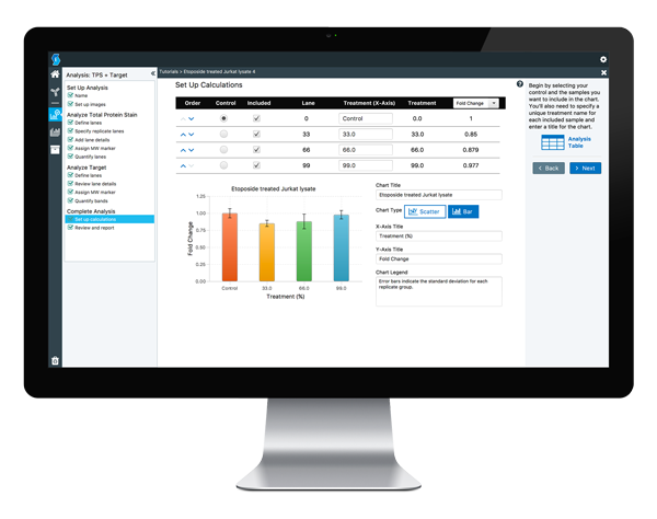
Pair your Odyssey CLx Imager with Image Studio™ Software for streamlined image acquisition and organization and with new Empiria Studio® Software for analysis of your Western blots.
Do more with consistent, robust digital imaging technology that has propelled the Odyssey imager family to over 10,000 peer-reviewed publications. See the benefits of stable NIR fluorescence and detect multiple targets in the same lane. Get the most accurate results with over 6 logs of linear dynamic range, so you can make more discoveries.
Need more information? Contact us.
* Remote demonstrations are available.