Study Cell Surface Receptors with On-Cell Westerns

On-Cell Western Assay

What It Is

An On-Cell Western (OCW) Assay is a cell-based assay that enables quantitative monitoring of cell surface protein expression. It can analyze multiple samples quickly and quantitatively, and it eliminates the use of radioactivity for the assay. The OCW Assay can be used to:

  • Detect and quantify target proteins localized to the cell surface
  • Quantify ligand binding to cell surface receptors
  • Monitor receptor internalization and recycling by detecting loss and reappearance at the cell surface
  • Perform and detect cell surface biotinylation assays
  • Evaluate the effects of mutations, targeted therapeutics, and other treatments on protein expression and trafficking

How It Works

With both standard In-Cell Western™ and OCW Assays, cells are first seeded in a microplate.

With In-Cell Western Assays, cell membranes are permeabilized after cells are treated and fixed, so antibodies can reach antigens inside the cell. (Not familiar with the In-Cell Western Assay? Learn more here.) However, the permeabilization process can compromise the detection of antigens present on the cell membrane.

With OCW Assays, cells are not permeabilized because the protein targets are detected on the cell surface—not intracellularly. Instead, they are stained with antibodies against extracellular protein domains, so only cell surface antigens are detected.

The cells in both assays can then be imaged either live or post-fixation.

Ways to Use It

Monitor Receptor Internalization and Recycling

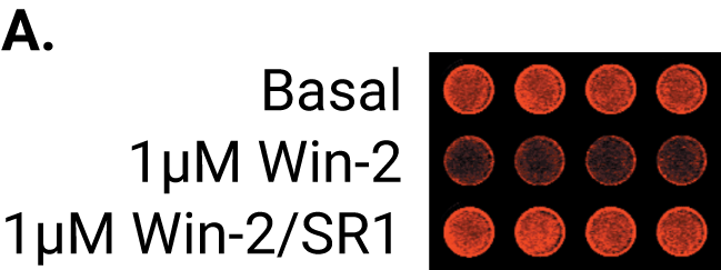
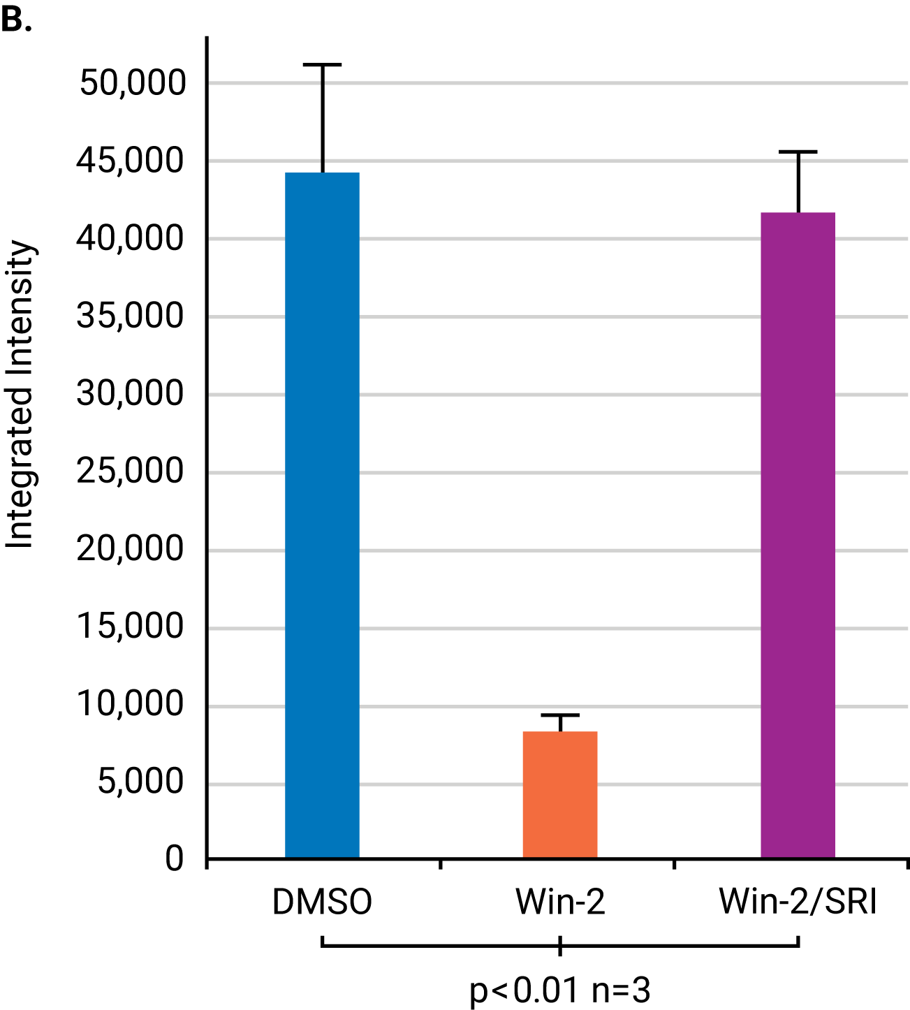
A study by Wager Miller used the OCW Assay to study the internalization and recycling of cannabinoid receptor 1 (CB1), a G protein-coupled receptor, after treatment with receptor agonists and cycloheximide. The antibodies were targeted against specific extracellular or intracellular domains of CB1. The observed time course for receptor internalization was consistent with previous confocal microscopy studies.

figure 1A
figure 1B
Figure 1. Effect of Win-2 agonist on CB1 internalization in HEK293 cells measured by an OCW Assay. A) Internalization of CB1 occurs after the addition of Win-2 agonist, as indicated by a decrease in integrated intensity. Treatment with SR1 antagonist recycled CB1 to the cell surface, as indicated by the restoration of the integrated intensity. Imaged on an Odyssey® Imager. B) Graphical representation of the integrated intensities from Figure 2A imaged on an Odyssey Imager. Reprinted with permission from Wager Miller.1

Quantify Plasmalemmal Expression of an Ion Channel

A study by Royal, Tinker, & Harmer investigated the roles of two phosphatidylinositols—Phosphatidylinositol-4,5-bisphosphate (PIP2) and Phosphatidylinositol-4-phosphate (PI(4)P)—in the anterograde trafficking of KCNQ channels. They used OCW and ICW Assays to determine the surface expression (Fig. 2A) and trafficking (Fig. 2B) of VSV-KCNE1-KCNQ1 (VSV-E1-Q1), a construct whose VSV epitope was used to monitor cell-surface expression of the KCNQ1-KCNE1 (Q1/E1) channel complex.

Overall, results indicate that trafficking and expression are not affected by PIP2 and/or PI(4)P reduction at the plasma membrane or by PI(4)P reduction at the Golgi. Consequently, the Q1/E1 channel may not require PIP2 or PI(4)P for anterograde trafficking. However, PIP2 may be necessary for the activation of the Q1/E1 channel once at the plasma membrane (PM).

figure 2
Figure 2. Channel cell-surface and total expression in Human Embryonic Kidney 293 (HEK293) cells were unaffected by the reduction of PIP2 and PI(4)P at the plasma membrane (PM) and PI(4)P at the Golgi. A) VSV-E1-Q1, LYN11-FRB, and either PJ-DEAD or PJ were transiently expressed in HEK293 cells. Rapamycin (Rap) was added to recruit the constructs to the PM. OCW results indicated that PJ-DEAD and PJ had no effect in the absence of rapamycin or PIP2/PI(4)P depletion at the PM. B) VSV-E1-Q1, Tgn38-FRB, and either PJ-DEAD or PJ-SAC were transiently expressed in HEK293 cells. Rap was added to recruit the constructs to the Golgi. OCW results indicated that PJ-DEAD and PJ-SAC had no effect on channel cell-surface and total expression in the absence of rapamycin or PI(4)P depletion at the Golgi.2

Confirm Membrane Expression of GPCR Constructs

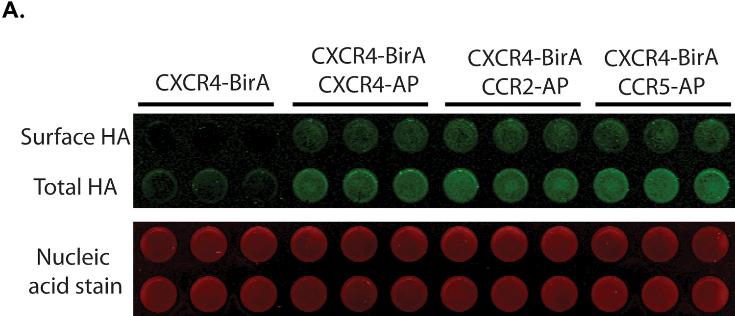
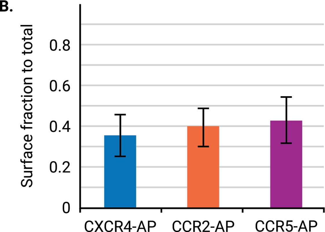
A study by Steel, Murray, and Liu examined the dimerization of G protein-coupled receptors (GPCRs), which help mediate human physiological responses by engaging with signaling pathways. To test a multiplex biotinylation assay, they used three chemokine receptors: CXCR4, CCR2, and CCR5. An OCW Assay was used to confirmed surface expression of the HA-tagged receptors (Fig 3). The proximity-based biotinylation assay confirmed CXCR4 homodimerization and CXCR4-CCR2 and CXCR4-CCR5 heterodimerization.

figure 3A
figure 3B
Figure 3. Surface expression of chemokine receptors using an OCW Assay. A) Cells were labeled with anti-HA. Fluorescence signals (green) were scanned then normalized to a nucleic acid stain (red). B) Surface expression of the chemokine receptors averaged 30 to 40 percent.3

References

  1. Wager-Miller, J. (2004). Tracking G Protein-coupled Receptor Trafficking Using Odyssey Imaging.
  2. Royal, A.A., Tinker, A., & Harmer, S.C. (2017). Phosphatidylinositol-4,5-Bisphosphate Is Required for KCNQ1/KCNE1 Channel Function but Not Anterograde Trafficking. PLoS ONE 12(10): e0186293. https://doi.org/10.1371/journal.pone.0186293
  3. Steel, E., Murray, V.L., & Liu, A.P. (2014). Multiplex Detection Of Homo- And Heterodimerization Of G Protein-Coupled Receptors By Proximity Biotinylation. PLoS ONE 9(4): e93646. https://doi.org/10.1371/journal.pone.0093646