See more than ever before.
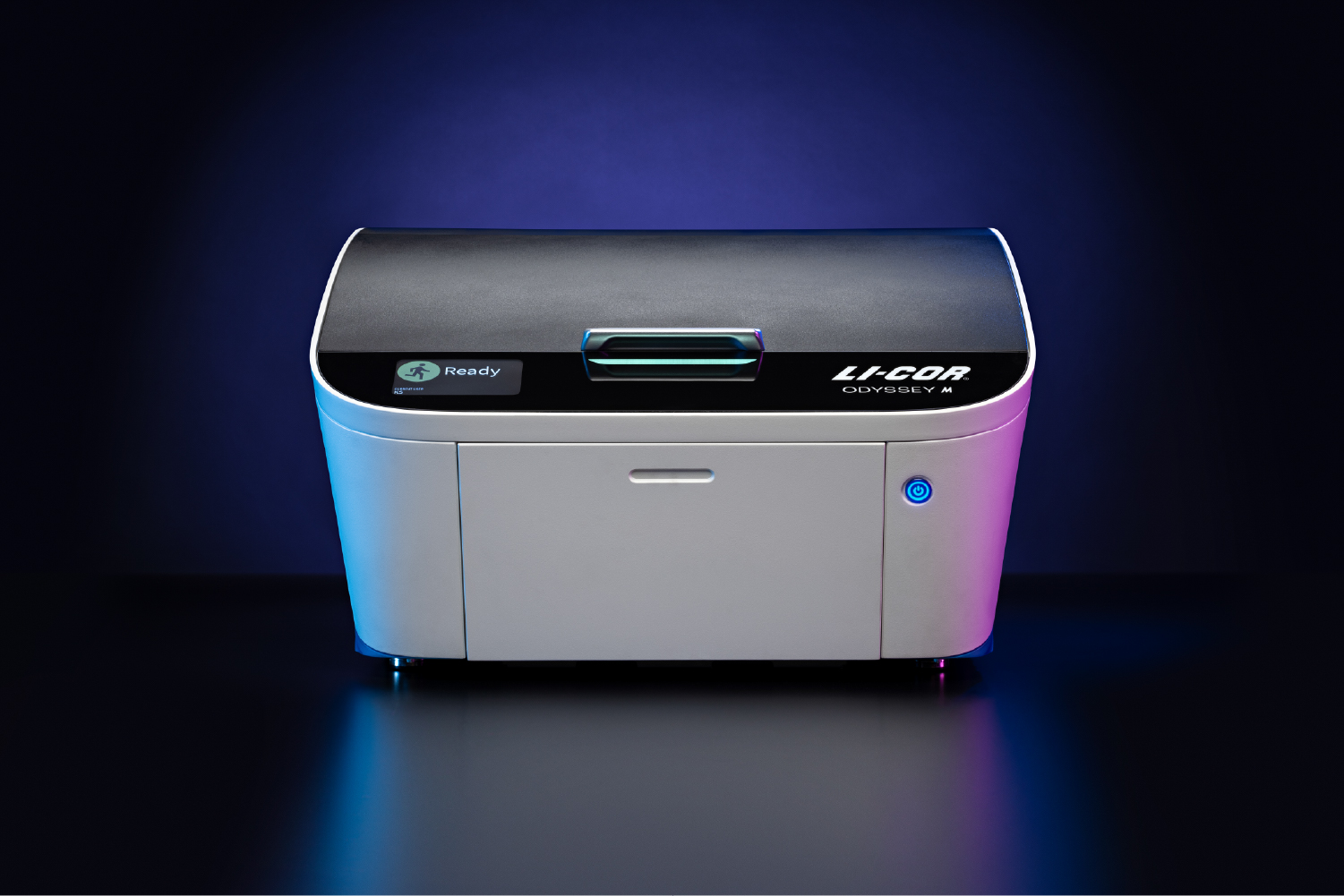
For more in-depth information from every sample scanned, we gave the Odyssey M a sophisticated sCMOS image sensor and industry-leading 5-micron resolution. No other imager can give you this level of sensitivity and dynamic range. With NIR and visible fluorescence, RGB, and optional luminescence capabilities—all 19 channels calibrated and integrated for consistent performance—you can easily image an ever-expanding variety of assays. And our innovative line-scanning technology leads the industry in speed. The Odyssey M acquires all this data in minutes, not hours.
The Odyssey M is truly unmatched in application versatility, performance, technology, and scientific expertise.
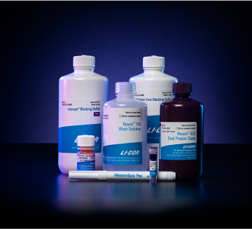
There are no expensive and delicate optical modules to purchase, manage, and assemble for different acquisitions.
One multimodal imager provides you and your lab with unmatched application versatility and data quality.
The unparalleled versatility of the Odyssey M makes it an optimal choice for any lab.
The industry’s most sophisticated imager enables you to make discoveries and decisions quickly with confidence.
From acquisition through analysis, quickly get the results you need to answer research questions–or to ask new ones.
For pricing on Odyssey M imagers or other LICORbio instruments, contact our Biotechnology Sales team.

 Powered by Bioz