D-DiGit Gel Scanner for Safe DNA Gel Imaging

D-DiGit® Gel Scanner

Complete nucleic acid gel analysis documentation without hassles and hazards.

Get a Quote
D-DiGit
D-DiGit with open lid

Safeguard your lab and research

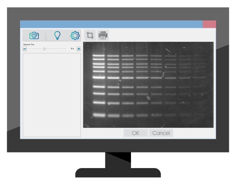
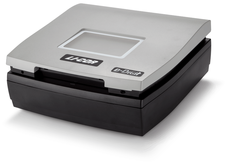
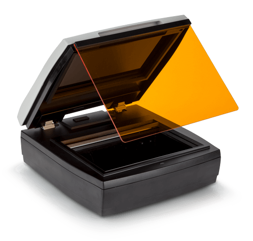
Perform your nucleic acid gel imaging with a safe, consistent, single-system workflow with the D-DiGit Gel Scanner.

UV-free imaging

Upgrade to safer, high-sensitivity LED detection that does not interfere with cloning and downstream processing of excised fragments. DNA fragments exposed to UV light during gel imaging and band excision could be nicked or damaged, resulting in lower cloning efficiencies1, 2, 3, 4, 5. Additionally, a non-UV light source eliminates the risk of accidental UV exposure during gel visualization and band excision.

“…researchers often assume that the damage caused by visualizing DNA on a UV transilluminator has little effect on subsequent DNA manipulations. However, several reports now make it clear that exposure of DNA can have a significant detrimental effect on downstream protocols involving cloning and biological activity.”

Paabo, S et al.2

Safer stain alternatives

The D-DiGit Gel Scanner enables safe and convenient detection and extraction of DNA and RNA samples stained with fluorescent safe stains, like SYBR® Safe. Visualize sample migration and conduct gel excision without contamination or the need to transfer gels between viewing and cutting services, and safely excise bands of interest to extract nucleic acids for downstream application. Safe stains are environmentally-friendly alternatives to ethidium bromide, and when added either during gel preparation, or as a post-electrophoresis stain, safe stains eliminate the need for hazardous disposal and hassles associated with ethidium bromide.

The D-DiGit detects a variety of safe stains that have an excitation maximum of approximately 490 nm and emission greater than 520 nm. See a list of compatible safe stains.

Streamline your workflow and improve protocol efficiency

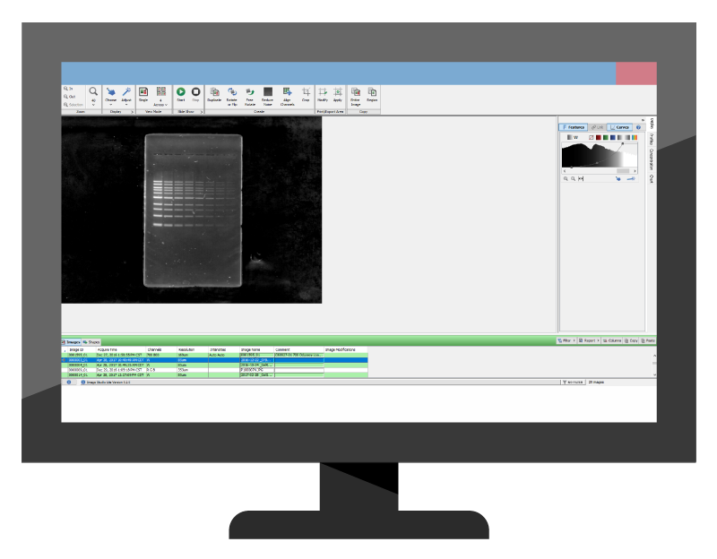
Get the most out of your gel imager. Scan gels, small and large; excise bands; document your work; and visually analyze data; all on a single, compact platform.

D-DiGit illustration

The D-DiGit Gel Scanner provides a large scan area to visualize most medium and larger format gels. Nucleic acid band extraction and imaging can be performed directly on the scan surface. It comes fully configured, so you don’t need to assemble parts, or transition between separate viewing and gel extraction platforms. Just place your gel on the scan area and perform your analysis. For convenience, a viewing shield is provided with the scanner.

Download the D-DiGit Image Acquisition Software at your workstation for easy image capture and archiving. With a user-friendly interface and a range of exposure times, imaging gels is quick and convenient. The D-DiGit software captures images that are compatible with LI-COR Image Studio and Image Studio Lite software for further data quantitation, analysis, and annotation.

gel viewing illustration
data imaging illustration
Gel viewing
Imaging
band excising illustration
data analysis illustration
Band excising
Analysis
Consolidate your nucleic acid gel analysis on a single system dedicated to high-sensitivity imaging. Use the D-DiGit acquisition software for gel viewing and imaging and use Image Studio software or free Image Studio Lite software for organization and analysis of your images.
D-DiGit data illustration

Get the complete picture with excellent sensitivity

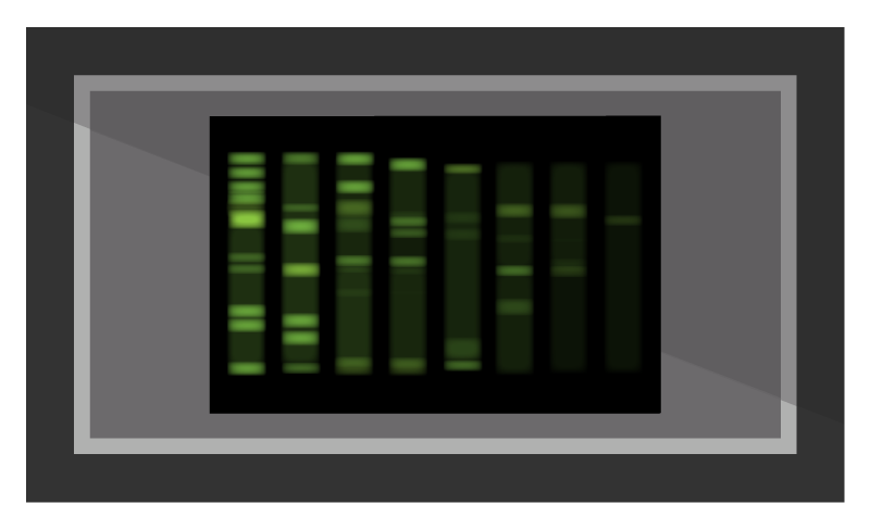
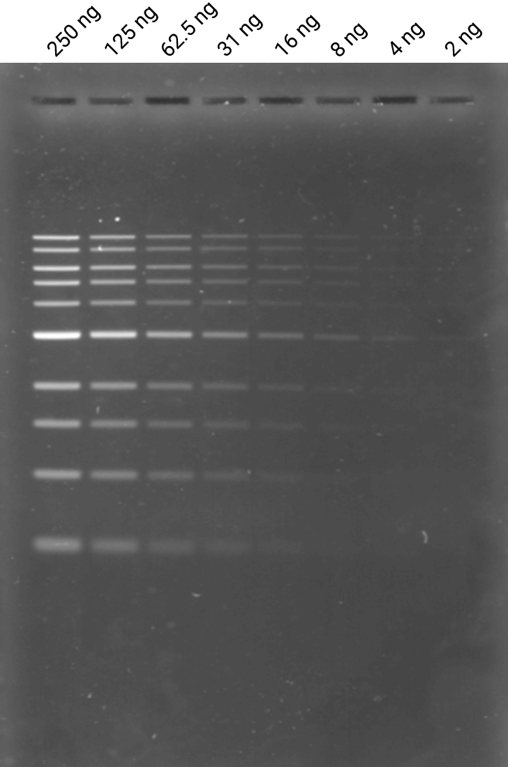
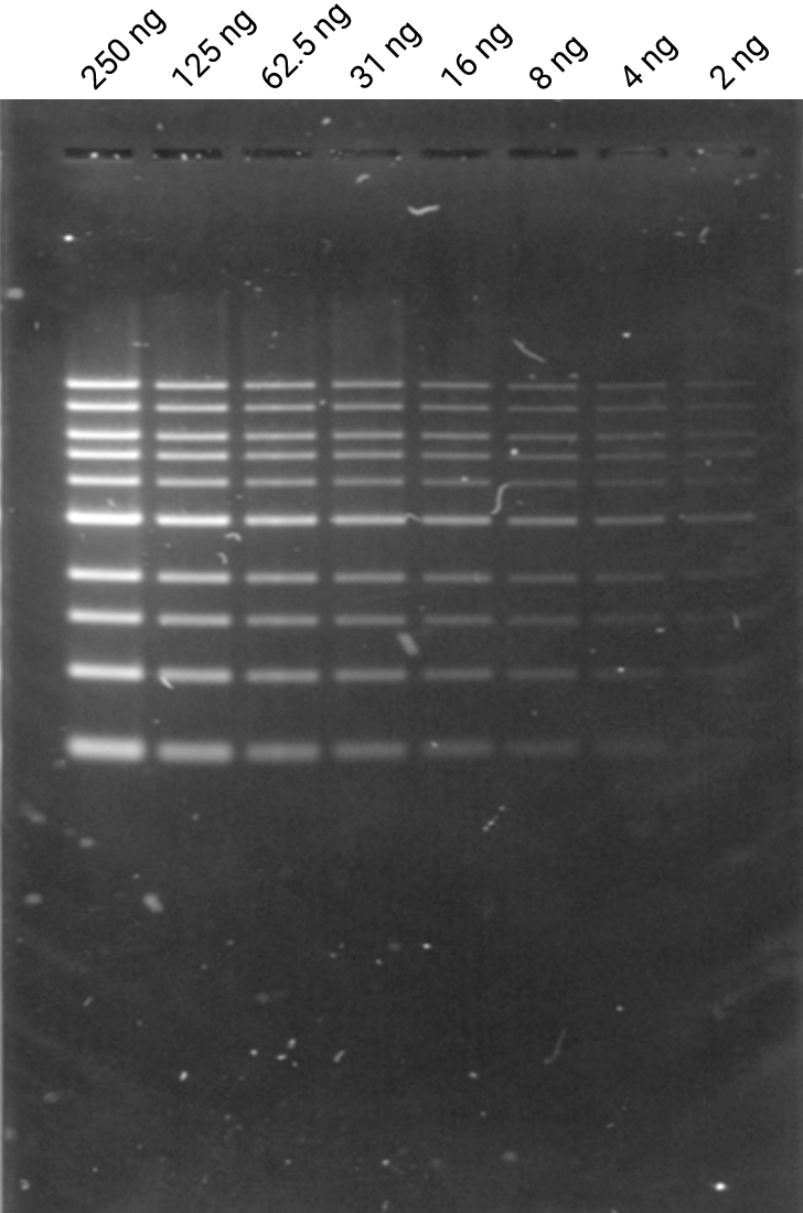
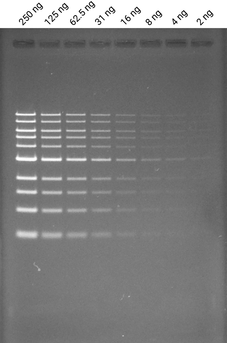
Get the complete picture without compromising data quality. Excellent sensitivity provided by the optical system and fluorescence chemistry lets you detect even minute quantities of sample with confidence.

Get excellent sensitivity to detect minute quantities of DNA and RNA on agarose gels. Engineered with a blue LED light source and high-resolution CCD detectors, the D-DiGit Gel Scanner detects as little as 0.04 ng of nucleic acid per band.

With a non-UV light source, you get safe and sensitive detection that does not interfere with downstream cloning applications. Safe stain imaging of nucleic acid gels on the D-DiGit Gel Scanner yields detection sensitivity comparable to ethidium bromide on a UV transilluminator.

GelRed data
A. Gel stained with 3X GelRed (Biotium)
SYBR data
B. Gel stained with 1x SYBR Safe (Invitrogen)
ethidium bromide data
C. Gel stained with 0.5 µg/ml ethidium bromide
A dilution series (1:2) from 250 ng to 2 ng of NEB 1kb DNA ladder was loaded in lanes 1-8 on three separate agarose gels and electrophoresed. Gels A, B, and C were stained post-electrophoresis. The D-DiGit was used to image gel A at 12X exposure setting, followed by gel B at 4X exposure setting. Gel C was imaged on UV transilluminator (UVP).
D-DiGit

Order now

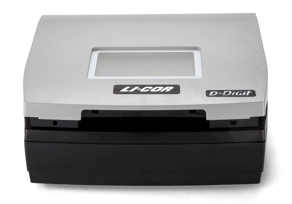
Perform your nucleic acid gel analysis with a safe, consistent, single-system workflow with the D-DiGit Gel Scanner.

Instrument

D-DiGit Gel Scanner

Quantity

Price

To order, contact your local distributor.

Need a quote? Get one now.

Need more information? Contact us.

Is your lab also performing chemiluminescent Western blot imaging?

Save money and bring digital scanning technology for both protein blot and nucleic acid gel imaging to your lab with the DiGit Duo bundle, featuring the D-DiGit Gel Scanner and C-DiGit® Blot Scanner. Learn more.

References
  1. Grundemann, D.; Schomig, E. BioTechniques 1996, 21, 898–903.
  2. Paabo, S.; Irwin, D. M.; Wilson, A. C. J. Biol. Chem. 1990, 265, 4718–4721.
  3. Hartman, P. S. BioTechniques 1991, 11, 747–748.
  4. Cariello, N. F.; et al. Nucleic Acids Res. 1988, 16, 4157.
  5. Hoffman, L. EPICENTRE Forum 1996, 3, 4–5.