Tissue section imaging provides insight into biological processes, such as disease onset and progression. Tissue sections also help scientists identify treatment targets and develop potential therapeutics.
To visualize targets at a subcellular level, you must examine tissue sections using microscopy. However, searching through each slide one at a time for areas of interest is challenging and time-consuming. It’s very much like looking for a needle in a haystack.
Whole slide imaging (WSI) can help you microscopically screen slides to find those of interest. While effective, traditional whole slide imaging requires a dedicated imager and a complex and costly system to manage the large image files.
Whole slide triage imaging (WSTI) is the latest advancement to quickly screen slides at the macroscopic level and determine which slides require closer inspection at the microscopic level. Think of it as a way to pull just the needles from the stack–and ignore all the hay.
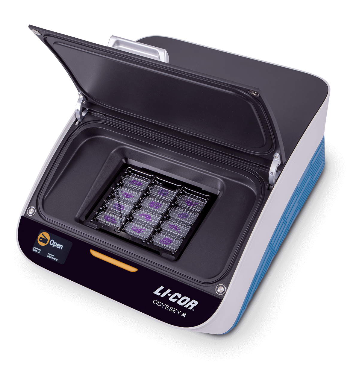
Whole slide triage imaging is a new application based on the unique features of the Odyssey® M Imager. This application addresses the shortcomings of screening tissue section slides using either microscopy or whole slide imaging.
| Whole Slide Imaging (WSI) | Odyssey M (WSTI) | ||
|---|---|---|---|
Time Taken per Acquisition |
H&E Slide | 2.5 min | 1.5 min |
| Two Fluorescent Targets | 8 min | 1.5 min | |
| Total Time for One Slide Pair | 10.5 min | 3.0 min | |
| 25 Total Slide Pairs (H&E + Fluorescence) | |||
| Total Time for 25 Slide Pair | 4.375 hrs | 1.25 hrs | |
With whole slide triage imaging, you can quickly screen slides before microscopic analysis by imaging multiple slides at a resolution of 5 microns–which is optimal for speed, clarity, and file size.
With the Odyssey M, you can also image thicker tissue sections. Imaging thicker samples limits the number of slides you need to screen and is useful for tissue sections that will not require subcellular resolution imaging.
| Whole Slide Imaging (WSI) | Odyssey M (WSTI) | ||
|---|---|---|---|
File Size per Acquisition |
H&E Slide | 2000 MB | 25 MB |
| Two Fluorescent Targets | 4000 MB | 50 MB | |
| Total Storage Space for One Slide Pair | 6000 MB | 75 MB | |
| 25 Total Slide Pairs (H&E + Fluorescence) | |||
| Total Storage Space for 25 Slide Pair | 150 GB | 1.88 GB | |
Additionally, your data management costs are significantly lowered by:
There are several features of the Odyssey M Imager that make it ideal for whole slide triage imaging. With the Odyssey M, you can:
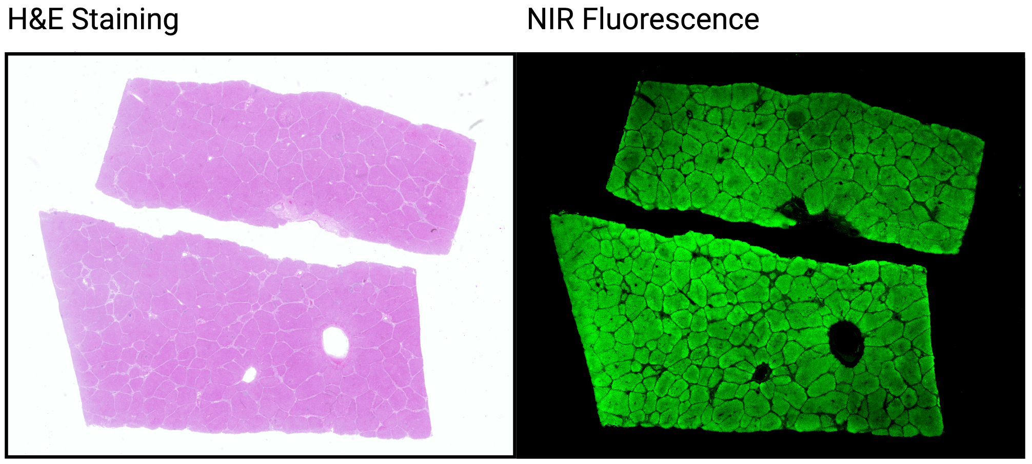
Since the Odyssey M images all slides using the same resolution, you can confirm observations by quickly and easily overlaying serial sections from multiple channels. First, observe diseased tissue in RGB true color with H&E stain on one slide. Then, assess biomarker location and localization using visible or near-infrared fluorescence on an adjacent slide.
Quickly narrow down the number of slides you need to take into microscopy. The Odyssey M performs fast image acquisitions and allows you to scan up to 12 slides at a time using thicker samples. Imaging thicker tissue sections at the beginning means you will have fewer slides to screen, which reduces the total amount of time spent imaging. Additionally, the wide dynamic range of the Odyssey M records the full range of data in one acquisition–there is no need for a second scan.

The wide dynamic range of the Odyssey M also improves consistency. Because you acquire all data in one scan, you eliminate any potential variability due to timing, positioning, or other factors. The included slide holder for the Odyssey M ensures you place your slides consistently for all acquisitions.
The 5-micron resolution of the Odyssey M is the optimal resolution to perform whole slide triage imaging. It is detailed enough to enable decision-making and may even eliminate the need for subcellular imaging depending on the application; yet, it allows for fast image acquisitions and file sizes that are much smaller and more manageable.
The unique characteristics of the Odyssey M Imager make whole slide triage imaging possible. The Odyssey M strikes a balance between acquisition speed, image quality, and file size to let you screen quickly while minimizing data management costs.
To start triaging your tissue section slides, visit the Odyssey M page.