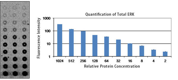
Protein arrays are a high-throughput way to generate information about protein abundance or modification state.
When you use near-infrared fluorescence to detect your protein arrays, you will get:
Protein arrays with spots that are equal to or greater than 85 μm are compatible with the Odyssey® DLx Imager, whereas spots that are equal to or greater than 500 μm are compatible with the Odyssey XF Imager.
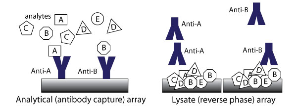
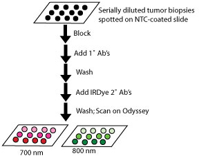
Lysate (reverse phase) arrays contain complex samples, such as cell or tissue lysates, that are printed on an array surface and interrogated with antibodies.4