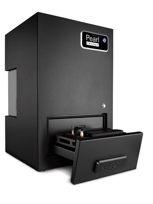
Pearl Trilogy Imaging System for in vivo Imaging Applications
Pearl Trilogy

Pearl® Trilogy
Small Animal Imaging System

Characterize and monitor therapeutic delivery and response using a sensitive, two-target detection platform.

Get a Quote
Pearl Trilogy

See More Details with Near-Infrared Fluorescence

Achieve high signal-to-noise ratios by minimizing tissue autofluorescence. Near-infrared (NIR) fluorescent imaging provides exceptional sensitivity for sharp detection of your targeted therapeutic, tumors, and other targets in live animals and excised tissue.
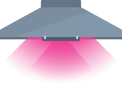
Optical Imaging in a New Light

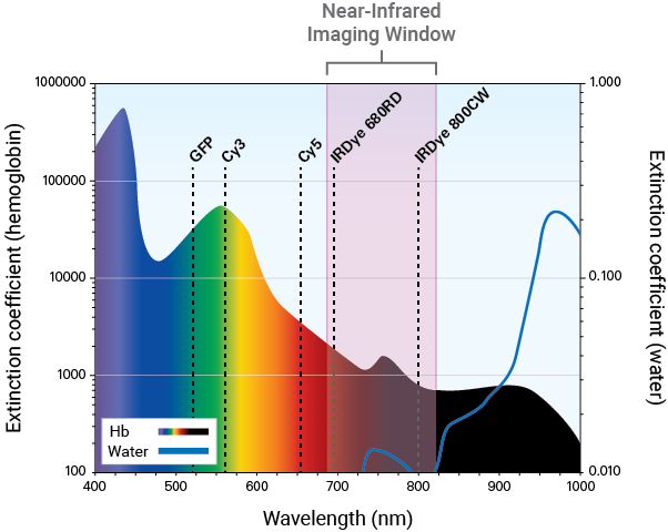
NIR fluorescent detection allows you to capture a detailed representation of the in vivo activity of your therapeutic. In the NIR spectrum (680 nm to 820 nm), autofluorescence from tissue is significantly reduced. As a result, animal tissue is rendered virtually transparent, allowing light to penetrate to targets located deep within the body.

NIR fluorescence provides exceptional sensitivity with high signal-to-noise ratios. High signal-to-noise ratios provide an accurate representation of therapeutic activity throughout the entire body of the animal. This level of detail enables you to evaluate specificity, biodistribution and clearance, and efficacy with accuracy and confidence.

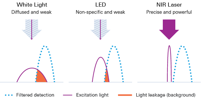
The Pearl Trilogy couples NIR lasers with filter technology to achieve a stable source of precise and powerful illumination. This leads to sharp excitation light within narrow wavelengths, causing minimal diffusion across the imaging field (CVs < 3%).

White light and LED light sources diffuse excitation light across the imaging field, causing variation in signal intensities and inconsistent data. Even with appropriate excitation and emission filters, some leakage of excitation light occurs, leading to high background.

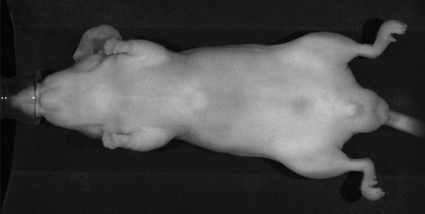
Image A

A. White Light

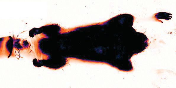
Image B

B. Visible

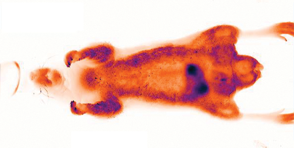
Image C

C. Near-Infrared

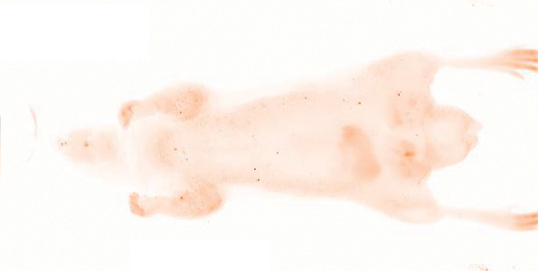
Image D

D. Near-Infrared

Tissue autofluorescence is much lower at NIR wavelengths. Tissue autofluorescence is much higher in white light (A) and visible light (B) than in NIR wavelengths (C, 700 nm, and D, 800 nm). Autofluorescence at 532 nm was very high.
light sources
Get the sensitivity and image quality you need to detect subtle changes. NIR lasers deliver powerful, precisely-tuned excitation light to generate strong signals and provide superior image quality allowing for detection of deeper targets and low tissue autofluorescence.

figure
Don’t let visible light sources limit what you can see. Capture images in the NIR window for optimal sensitivity.

Take Advantage of Multitarget Detection

Use the Pearl Trilogy to evaluate the specificity and delivery system of your targeted therapeutic. Two-color imaging allows you to visualize colocalization to develop therapeutic strategies and to confirm your therapeutic reaches its intended target.
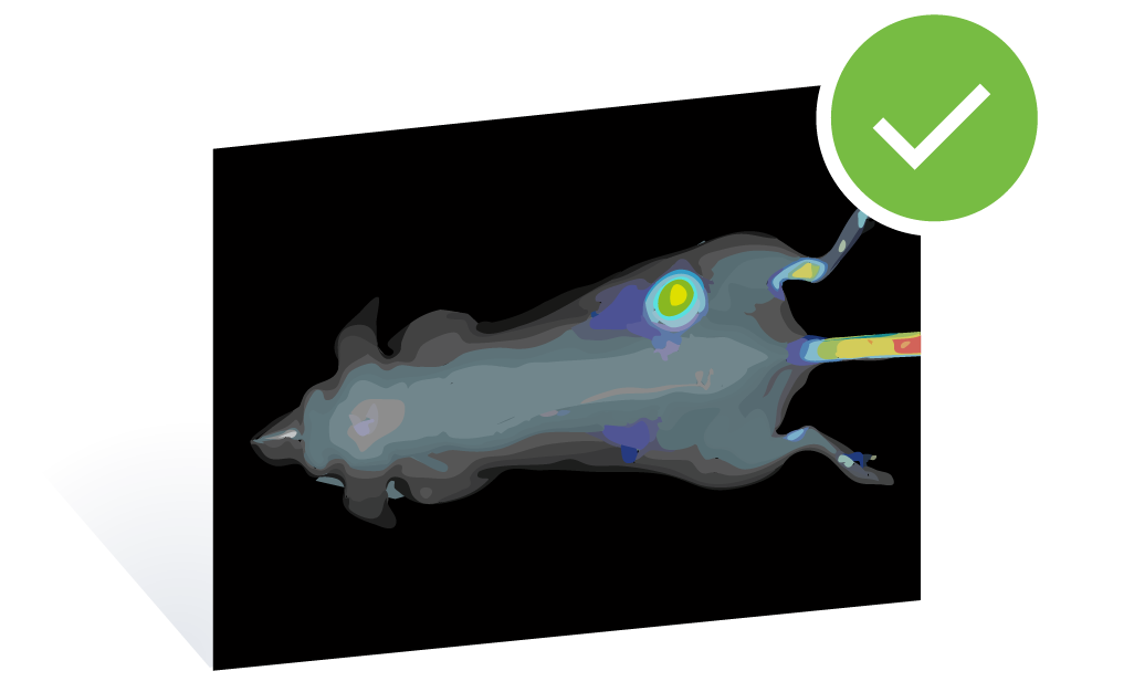
Best Shot, Everytime

Colocalization of targets can be used to identify disease risk factors, to reveal potential targets for therapeutic treatment, and to assess specificity and delivery. With multicolor NIR fluorescence imaging on the Pearl Trilogy, you can label two separate targets using spectrally distinct dyes. Those targets can then be detected simultaneously using the 700 nm and 800 nm channels, and the images can be overlaid to examine colocalization.

figure
Imaging of multiple targets in the same animal. IRDye® 680 BoneTag™ agent reveals skeletal features (700 nm, grayscale). IRDye 800CW EGF (800 nm, pseudocolor) was used to image an A431 tumor on the right hip. Overlay shows combined 700 nm and 800 nm images.

Minimize Variability in Your Data

Pearl Trilogy imagers are engineered to deliver high-quality and consistent data image after image while simplifying repeated imaging. This lets you accurately profile biodistribution and clearance and detect subtle changes in tumor size over time to characterize therapeutic response and efficacy.
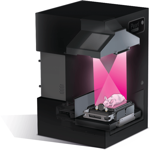
Some experiments, such as longitudinal studies, require moving animals in and out of the imaging area. The FieldBrite™ Xi2 system in Pearl Imagers is designed to provide uniform illumination for every image. Whether your experiment requires repeated imaging of one mouse or twenty mice, the camera settings are consistent for each capture—without needing readjustment.

Pearl Trilogy animation

Have Complete Confidence in Your Results

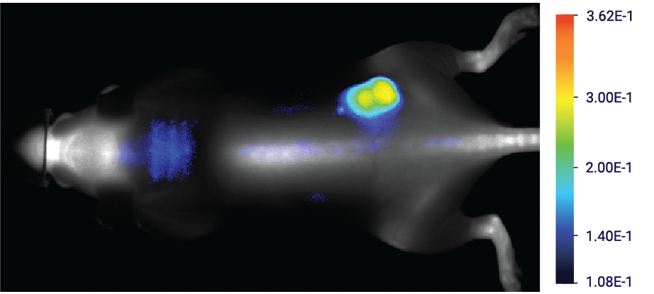
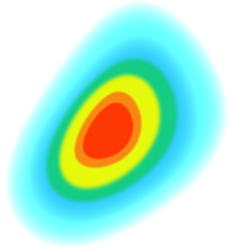
The Pearl Trilogy enables you to capture the full range of signal intensity—without saturation—in one image. Track both strong and faint signals to visualize a range of disease states in vivo and assess uptake and localization ex vivo using excised organs.
Full Range of Signal Intensity

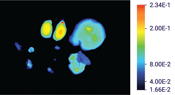
Imagers with limited dynamic ranges are unable to simultaneously detect signals differing more than 3 or 4 logs (1000 to 10,000X). As a result, images are saturated in areas of high signal intensity, and data are no longer coherent. Correction usually involves manual adjustment of camera exposure before every image capture.

The FieldBrite Xi2 technology in Pearl Trilogy imagers offers an industry-leading dynamic range of more than 6 logs. This wide-dynamic range provides a complete picture for accurate therapeutic characterization. Confidently measure variations in signal corresponding to biodistribution and clearance, response, localization, and other key factors for therapeutic development without needing to adjust any settings.

"Ease of use is definitely a very positive attribute. The fact that you can walk up to it and get in and get out is a real advantage…"

Dr. H Charles Manning, Vanderbilt University
Signals in tissue samples
Imaging of both strong and weak signals in excised tissue samples. Multiple organs may be imaged together to examine the uptake and localization of a targeted therapeutic. Imaging of low-signal targets, such as lymph nodes, alongside high-signal organs, such as kidneys, require a wide dynamic range.

Acquisition and analysis software for the Pearl Trilogy

Pair your Pearl Trilogy with LI-COR Acquisition Software and Empiria Studio® Software for advanced acquisition and analysis using intuitive tools.
data illustration
LI-COR Acquisition Software

LI-COR Acquisition Software

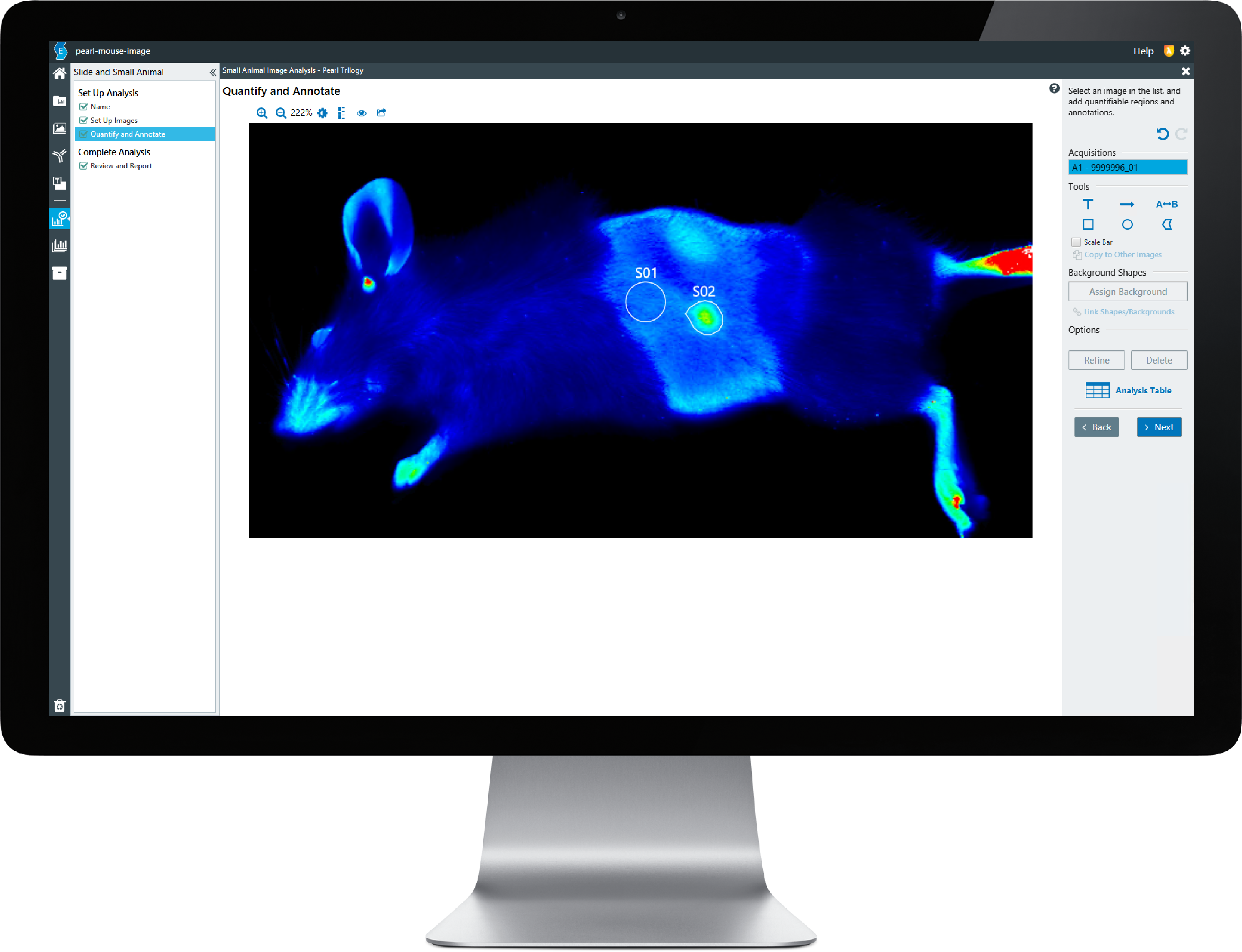
LI-COR Acquisition Software allows you to acquire, organize, and adjust images. A workflow-based system ensures no steps get missed, and you can acquire images using identical settings–every time. You can then export images for analysis in Empiria Studio or publication.

Empiria Studio

Empiria Studio Software

Empiria Studio enables you to perform fast analysis of your small animal images. At each step, Empiria Studio provides intuitive tools to quantify, examine, organize, and annotate images acquired on your Pearl Trilogy Small Animal Imager.

Learn More about Empiria Studio

Pearl Trilogy

Ready to Take the Next Step?

With the Pearl Trilogy, you can accomplish in-depth delivery and response studies backed by reliable in vivo and ex vivo data. Be confident you have fully characterized and validated your therapeutic in live animals to prepare for the next steps.

Need more information? Contact us.