Quick Western Kit
Components
Components for the Quick Western Kit (926-69100):
- IRDye® 680RD Detection Reagent
- Intercept® (PBS) Blocking Buffer
Other Required Reagents
- 1X PBS
- Primary Antibody
- 1X PBS-T (1X PBS containing 0.1% Tween® 20)
- 20% Tween 20
- 20% SDS (If using PVDF membrane)
Specifications
- Fluorophore: IRDye 680RD
- Excitation Wavelength: 676 nm (in PBS)
- Emission Wavelength: 694 nm (in PBS)
- Form of Detection Reagent: IRDye 680RD Detection Reagent, lyophilized in water. Contains 10 mg BSA (IgG and protease free) per mg of Detection Reagent as a stabilizer and 0.01% sodium azide as a preservative.
- Storage: Store at 4 °C and protect from light. Will remain stable for up to 3 months at 4 °C in reconstituted form.
- Kit will perform approximately 25 Western blots.
Contains sodium azide.
Applications
The Quick Western Kit – IRDye 680RD provides a universal antibody detection reagent that can be combined with the primary antibody incubation step, eliminating the need for a secondary antibody. The overall time to complete a Western blot is reduced, while providing the advantages of near-infrared detection. The kit can be used to detect primary antibodies from a variety of hosts and has been shown to recognize primary antibodies to recombinant tagged proteins (e.g. 6X His, Myc, FLAG, etc.).
The kit also serves as a detection solution for post-immunoprecipitation samples by Western blot because it does not bind to denatured mouse monoclonal or rabbit monoclonal antibodies. The key benefit is the ability to use the same antibody for immunoprecipitation and post-immunoprecipitation detection by Western blot.
The IRDye 680RD Detection Reagent recognizes denatured polyclonal antibodies and is not recommended for detection of samples that have been immunoprecipitated using polyclonal antibody.
Purity and Specificity
IRDye 680RD Detection Reagent is known to have high affinity for IgG from human, mouse, rabbit, guinea pig, goat, sheep, pig, cow, cat, dog, and donkey. The Detection Reagent is known to have lower affinity for mouse IgG1, mouse IgA, rat, horse, and hamster IgGs and will not detect primary antibodies from a chicken host. The Detection Reagent has been specifically tested and qualified for Western blot applications. If additional affinity is required, please use IRDye conjugated secondary antibodies for detection.
Reconstitution
Reconstitute contents of vial with 0.25 mL 1X PBS. Mix gently by inverting. Protect from light and allow to rehydrate for at least 60 minutes before use. Centrifuge product if solution is not completely transparent after standing at room temperature.
Western Detection Protocol - Quick Western Kit
Step 1. Wet Membrane
For Nitrocellulose Membranes
Wet in 1X
For Immobilon®-FL PVDF Membranes
- Wet for 30 seconds in 100% methanol.
- Wet in 1X
Step 2. Block the Membrane
Place the membrane in an incubation box and block the membrane with Intercept® Blocking Buffer for 1 hour at room temperature with gentle shaking.
Be sure to use sufficient blocking buffer to cover the membrane (a minimum of 0.4 mL/cm2 is suggested).
Step 3. Prepare Detection Solution
- Add primary antibody (dilute as recommended by vendor) to Intercept Blocking Buffer.
- Add the IRDye 680RD Detection Reagent to the primary antibody + Intercept Blocking Buffer solution. Use 1 µL of IRDye 680RD to every 1 mL of Intercept Blocking Buffer (1:1000 dilution).
- Add detergent:
- For nitrocellulose membranes use 0.2% Tween® 20 in the detection solution.
- For PVDF membranes use 0.2% Tween 20 and 0.02% SDS in the detection solution.
Optimization is required to determine the final concentration of detergents. Concentration can range from 0.1 - 0.2% Tween 20 and 0.02 - 0.1% SDS.
Step 4. Decant and Discard Blocker
Step 5. Add Detection Solution
Add the detection solution prepared in Step 3.
Step 6. Incubate Blot in Detection Solution
Protect from light during incubation.
- Incubate using your standard primary antibody incubation time at room temperature on a platform shaker.
- If the standard primary antibody incubation time is overnight, store the blot at 4 °C on a platform shaker.
Step 7. Wash Membrane
- Carefully decant detection solution.
- Rinse the membrane in 15 mL of 1X PBS-T (1X PBS + 0.1% Tween 20).
- Decant.
- Add 15 mL of 1X PBS-T and incubate for 5 minutes at room temperature with gentle shaking. Protect from light.
- Decant wash solution.
- Repeat 2 additional times.
Step 8. Rinse Membrane
Rinse membrane with 15 mL of 1X PBS.
Step 9. Scan Membrane
Protect the membrane from light prior to scanning.
Scan the membrane (wet or dry) on an Odyssey Imaging System using the 700 nm channel.
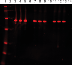
Example