Confirm Target Gene Knockdown or Knockout with Fluorescent RNAi Studies

Gene Knockdown and Knockout Confirmation

Knockdown and knockout are two methods used to silence a target gene of interest. The partial (knockdown) or complete (knockout) silencing of a target gene enables researchers to identify and validate biological targets of human health significance. These techniques also enable researchers to better understand disease processes, create negative controls, and advance therapeutic discovery.

Quantitative Western blotting and the In-Cell Western™ Assay can be used to confirm the effects of gene knockdown/knockout. Both assays can be performed with exceptional quality and consistency using Odyssey® Imagers, such as the Odyssey M.

Confirming Target Knockdown with RNAi Analysis

RNA interference (RNAi) is a naturally occurring process to inhibit, or knockdown, the normal expression of a gene by deactivating or suppressing mRNA. RNAi can be artificially replicated in the laboratory using an RNAi vehicle, such as siRNA, shRNA, or other RNA-producing vector constructs. This allows for experimentation with a gene of interest. Knockdown of a specific gene allows researchers to analyze genetic function or to identify targets with potential therapeutic applications.

RNAi Analysis Using Quantitative Western Blotting

Quantitative Western blots are an essential tool for RNAi analysis. With quantitative Western blots, you can:

  • Compare data from different vectors and constructs, such as siRNA, shRNA, or other RNA-producing vector construct
  • Confirm the impact of your RNAi strategy on protein levels
  • Analyze proteins in different signaling pathways to determine the impact silencing has on a phenotype, such as cell proliferation or migration

figure 1a
figure 1b
Figure 1. siRNA knockdown in HeLa cells was confirmed using an In-Cell Western Assay and Western blot. HeLa cells were transfected with no RNA (control), non-sense RNA (NS), or various amounts of AKT siRNA. Detection was performed using an AKT Rabbit primary antibody and IRDye® 800CW Goat anti-Rabbit Secondary Antibody with signals normalized to CellTag™ 700 Stain (In-Cell Western) and Revert™ 700 Total Protein Stain (Western bot). All images were acquired using an Odyssey® M Imager.

RNAi Analysis Using In-Cell Western Assay

In-Cell Western Assays can be used in a functional siRNA screen to measure knockdown effects in cultured cells. In-Cell Western Assays offer higher throughput for complex studies, as well as exceptionally consistent data (Z' factor).1

figure 2a
figure 2b
Figure 2. Schematic of the workflow for an siRNA screen. Adapted from Greg Hoffman, Harvard.

Hoffmann, et al. have demonstrated the In-Cell Western Assay can be used as a powerful cellular assay for genome-wide RNAi screens.2 In-Cell Western screening was used to assess the effects of knockdowns on mTORC1-dependent phosphorylation of ribosomal protein S6 (rpS6).2 Their research showed In-Cell Western RNAi screening to be faster and less expensive than high-content immunofluorescent microscopy while providing similar or better statistical replicability.

figure 3a
figure 3b
Figure 3. Results of pilot small molecule screen performed with a library of ~2500 known bioactive compounds. (A) Compounds with average Z-score < -2 are considered hits and are shown in red. Known inhibitors of mTORC1 signaling found in the library are shown in green. Star-shaped symbols represent compounds with > 4-fold reduction in cell number. (B) Plate to plate consistency is shown for a representative plate from the small molecule library.
figure 4a
figure 4b
Figure 4. In-Cell Western RNAi screening was faster and less expensive IF microscopy while providing similar or better statistical replicability. A side-by-side comparison of the In-Cell Western Assay and IF microscopy reveals that the In-Cell Western requires fewer reagents with significantly faster image acquisition and analysis. Additionally, the Z’factor, which measures assay reliability, was higher for the In-Cell Western Assay.

Confirming Target Knockout with CRISPR Analysis

CRISPR editing can be used to knockout a gene to inhibit the expression of certain proteins. Unlike knockdown, which inhibits a gene, knockout inactivates or removes a target gene completely. Like knockdown, knockout can be used to study the behavior and effects of a gene of interest to assess its function or its potential to affect human health.

CRISPR Analysis Using Quantitative Western Blotting

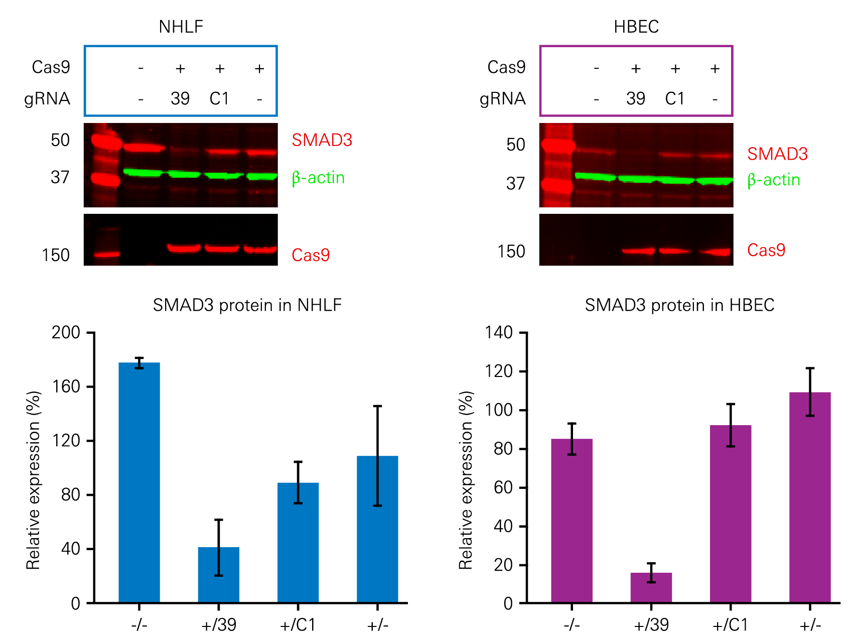
The effects of gene knockout via CRISPR can be confirmed using quantitative Western blotting. Figure 5 demonstrates the use of Adenoviral (AdV) CRISPR/Cas9 to inhibit the expression of SMAD3, a protein involved in triggering pulmonary fibrosis.

figure 5
Figure 5. AdV CRISPR/Cas9 exhibited significant SMAD3 knockdown in human primary cells. SMAD3 (red) and Cas9 (red) were detected in normal human lung fibroblasts (NHLF) and human bronchial epithelial cells (HBEC) and normalized to β-actin (green) using IRDye® Secondary Antibodies on near-infrared fluorescent Western blots. The presence of Cas9 and the gRNA 39 resulted in a significant decrease in SMAD3 protein. Images were acquired on an Odyssey CLx Imager. Adapted from Voets, O., et al. (CC BY).

CRISPR Analysis Using In-Cell Western Assay

The In-Cell Western Assay can be used to measure the effect of knockout in cultured cells. Figure 6 shows the efficiency of ERK1 knockout when compared to wild-type HeLa cells.

figure 6a
figure 6b
Figure 6. Knockout of ERK1 resulted in significantly lower expression versus the wild type. Wild-type and ERK1 knockout (KO 1, KO 2, and KO 3) HeLa cells were incubated with pan-ERK1 primary antibodies. CellTag™ 520 Stain for normalization and IRDye 680RD Goat anti-Mouse Secondary Antibodies were then added to the wells and the plate was imaged using the 520 and 700 nm channels of an Odyssey M Imager.

What steps can you take today toward improved knockdown/knockout confirmation?

LI-COR provides products, protocols, and support for Western blotting, In-Cell Western Assays, and many other assays that help reduce variability and increase confidence in your results. Want to learn more? Contact LI-COR today and let us help you improve your knockdown/knockout confirmation.

References

  1. Boveia, V., Ambroz, K.L.H., and Olive, D.M. (2009). Using the Z’-Factor Coefficient to Monitor Quality of Near-Infrared Fluorescent Cell-Based Assays. LI-COR Biosciences.
  2. Hoffmann, G., et al. (2008). ). A functional siRNA screen for novel regulators of mTORC1 signaling. Poster presented at ASCB Annual Meeting.
  3. Voets, O., Tielen, F., Elstak, E., Benschop, J., Grimbergen, M., Stallen, J., et al. (2017). Highly efficient gene inactivation by adenoviral CRISPR/Cas9 in human primary cells. PLoS ONE, 12(8). e0182974. DOI: 10.1371/journal.pone.0182974