Multiplexed Glycoprotein Staining with Odyssey Imagers
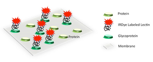
Glycoprotein Staining
With two-color near-infrared detection, the Odyssey® Imager offers a variety of applications for glycoprotein detection with multiplexing capabilities.
Near-infrared wavelengths provide low background from biological materials, buffer components, and standard membranes used in Western blotting and lectin blotting applications. The Odyssey Imager and IRDye® dye-labeled conjugates provide a single optimized solution for detecting a variety of glycoprotein interactions.1
Total Protein Detection for Glycoprotein Digestions (Coomassie staining)
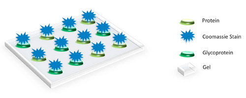
Figure 1. Schematic of total protein detection using Coomassie stain.
Visualization of all protein bands is possible with Coomassie staining. Glycoprotein bands with a known molecular weight can be identified by referencing a molecular weight marker lane on the gel or membrane. Phenomena such as shift changes due to denaturations or digestions can be documented when comparing treated to non-treated samples.
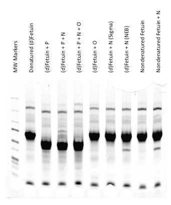
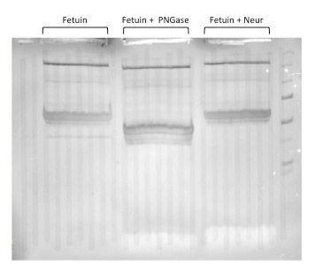
Figure 2. Example of glycoprotein detection with Coomassie on gels. Fetuin was digested with various glycoprotein specific enzymes, PNGaseF (P), neuraminidase (N), and O-glycanase (O). The reactions were separated on a 7.5% NEXT Gel and stained with Coomassie. The total protein stain of the gel confirms the PNGase F activity due to the protein shift in molecular weight. Neuraminidase and O-glycanase activities were confirmed using lectin-specific Western blots. Figure 3. Example of glycoprotein detection with Coomassie membrane staining. Fetuin was digested with either PNGase F or Neuraminidase and separated on a 10% NEXT Gel using a preparative comb. The proteins were transferred to Odyssey Nitrocellulose (P/N 926-31090), the membrane was stained with Coomassie and bands were detected using an Odyssey Imager. The image shows the protein band shifts to a lower molecular weight after digestion with PNGase F (largest shift) or Neuraminidase (smaller shift). Total protein detection can also be done on the same blot after detection with IRDye Infrared Dye probes.
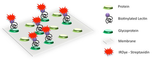
Indirect Detection using IRDye Labeled Streptavidin
Biotinylated lectins are commercially-available from several vendors and can be used in combination with IRDye Streptavidin to detect glycoproteins.
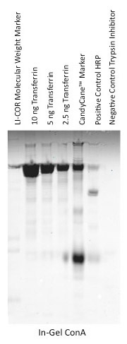
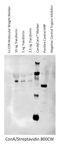
Figure 4. Indirect glycoprotein detection using IRDye Streptavidin. Transferrin contains N-linked glycoprotein structures and was separated on two reducing and denaturing 10% Bis-Tris gels. One gel was probed with biotinylated ConA and detected with IRDye 800CW streptavidin. The positive control was HRP (Horseradish Peroxidase). A second gel was transferred to nitrocellulose, probed with biotinylated ConA and detected with IRDye 800CW streptavidin. Both the gel and the membrane were imaged on an Odyssey Imager and showed signal with the lectin, ConA. This confirmed the presence of mannose residues, indicative of N-linked glycan structures. The positive control was horseradish peroxidase.
Direct Detection with an IRDye Labeled Lectin
Lectins can be covalently labeled with IRDye infrared dyes using an IRDye Protein Labeling kit. Labeled lectins can used to detect glycoproteins directly.
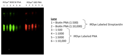
Figure 5. Direct glycoprotein detection using IRDye dye-labeled lectin. Fetuin was digested with neuraminidase to expose the PNA binding site and resolved on a 10% NEXT Gel (Amresco) using a preparative comb. Following transfer to nitrocellulose, the membrane was placed in the MPX™ Blotting System (P/N 921-00000). Each preparative well was then split into 6 individual MPX channels. PNA conjugates had previously been prepared using LI-COR labeling kits. The MPX channels were incubated with various dilutions of IRDye labeled PNA conjugates (Lane 3: 1:500; Lane 4: 1:1000; Lane 5: 1:5000; Lane 6: 1:10,000) and compared with IRDye biotinylated PNA (Lane 1: biotinylated PNA 1:5000; Lane 2: biotinylated PNA 1:10,000) and detected with IRDye Streptavidin at 1:1000 dilution.
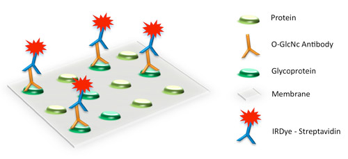
Glycoprotein Antibody Detection
O-GlcNAc (O-linked N-acetylglucosamine) modifications can be detected using a specific primary antibody and IRDye secondary antibodies.
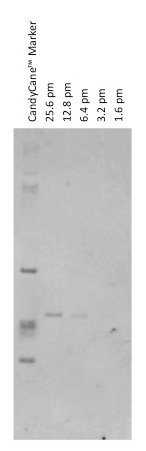
Figure 6. Glycoprotein detection using IRDye secondary antibodies. α-crystallin was detected using O-GlycNAc mouse monoclonal antibody and a IRDye 800CW Goat anti-mouse secondary. The blot was imaged on the Odyssey Imaging System.
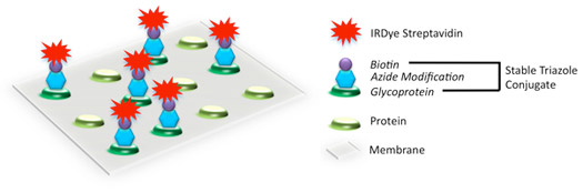
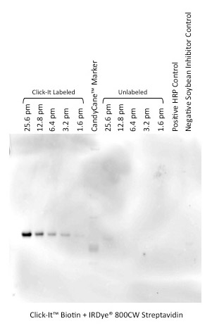
Enzymatic Glycoprotein Detection
O-linked glycosylated proteins can be modified with the Click-iT™ O-GlcNAc enzymatic labeling kit and detected using the Click-IT Biotin Glycoprotein Detection Kit in combination with IRDye 800CW Streptavidin.
Figure 7. α-Crystallin labeled via Click-iT chemistry using enzymatic O-GlcNAc labeling kit. The modified and unmodified products were resolved on a gel and transferred to a nitrocellulose membrane. The IRDye 800CW Streptavidin (P/N 926-32230) was used to detect the modified α-crystallin product by Western blot.
Typical Glycan Structures
Glycosylation is an area of post-translational protein modification receiving more attention by researchers as potential cancer biomarkers. Attachment of the sugar chain to the protein determines whether the interaction is deemed N- or O-glycosylation. These interactions of the glycoprotein structure can be represented by symbol nomenclature. Typical glycan structures are shown here.