Detect Proteins Directly with In-Gel Westerns

In-Gel Westerns

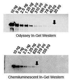
In-Gel Westerns directly detect protein in the polyacrylamide gel, without membrane transfer or blocking. Near-infrared (NIR) fluorescent In-Gel Westerns can be imaged with the Odyssey® M or Odyssey DLx Imagers when using IRDye® secondary antibodies for detection.

figure 1
Figure 1. Multiplex detection of two target proteins by In-Gel Western. Two-fold dilutions of a T7-Tag extract (red; 700 nm) and purified Transferrin (green; 800 nm) were detected using the In-Gel Western protocol and IRDye secondary antibodies. Gel was imaged using the Odyssey imaging system. Reprinted from Urh, M. et al. Poster presentation. Advances in Genome Biology and Technology Conference (2002).

In-Gel Westerns are useful for:

  • Large, hydrophobic, or post-translationally-modified proteins, such as glycoproteins, receptors, or cell membrane proteins that do not transfer well
  • Small proteins that may pass through the membrane during transfer
  • Multiplexed, two-color detection of two different protein targets within the same gel

Advantages of Near-Infrared In-Gel Westerns

  • Save time and reduce cost of running a blot
  • Eliminate variables associated with membrane transfer and blocking
  • NIR In-Gel Westerns provide sensitivity comparable to chemiluminescent In-Gel detection with clearer, sharper bands (Figure 2), but not as high as traditional NIR membrane Western detection.
  • Detect to nanogram to picogram levels (Figure 3)
  • Produce clean images with minimal background banding
figure 2
Figure 2. Sensitivity of Odyssey infrared In-Gel Westerns is equal to or better than chemiluminescence. Beginning with 10 ng/lane (far left), two-fold serial dilutions of purified Transferrin were separated by electrophoresis on duplicate gels. In-Gel Westerns were detected with infrared fluorescence (top) and chemiluminescence on film (bottom). Odyssey detection outperformed chemiluminescence.
figure 3
Figure 3. In-Gel detection of Cytochrome P450 3A4 (CYP3A4). Fixed gel was probed with anti-CYP3A4 primary antibody and IRDye 800 secondary antibody. The limit of detection is approximately 3 ng. Reprinted with permission from Theisen, M. J. and Chiu, M. L. LI-COR Biosciences (2004)1

Typical In-Gel Western Workflow

Perform electrophoresis

step 1

Remove stacking gel

step 2

Add fixation solution to fix proteins in gel

step 3

Wash

step 4

Incubate with primary antibody

step 5

Wash

step 6

Incubate with secondary antibody

step 7

Wash

step 8

Excite with infrared lasers

step 9

Acquire image using Odyssey Imager and analyze

step 10

References

  1. Theisen, M. J. and Chiu, M. L. In-gel immunochemical detection of proteins that transfer poorly to membranes.