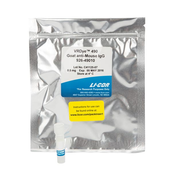
VRDye™ 490 Goat anti-Mouse IgG Secondary Antibody
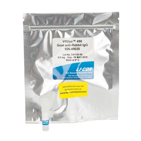
VRDye™ 490 Goat anti-Rabbit IgG Secondary Antibody
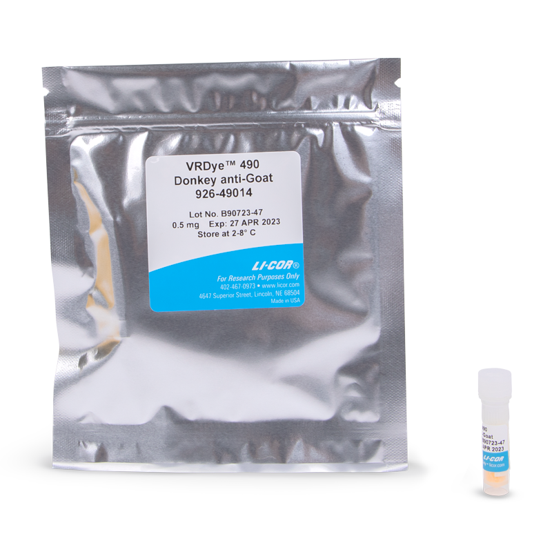
VRDye™ 490 Donkey anti-Goat IgG Secondary Antibody
VRDye™ 490 Donkey anti-Mouse IgG Secondary Antibody
VRDye™ 490 Donkey anti-Rabbit IgG Secondary Antibody
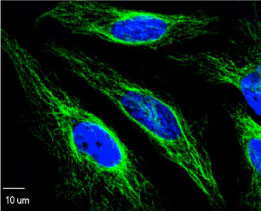
VRDye 490 secondary antibodies can be used for a variety of applications, including flow cytometry, immunofluorescence microscopy, and others.
These secondary antibodies are suitable for multiplex experiments when combined with other secondary antibodies labeled with proper fluorescent dyes and using instrumentation with appropriate excitation and detection capabilities.
Visible Fluorescence Reagents Overview
VRDye 490 secondary antibodies are supplied as purified immunoglobulin conjugates, lyophilized in phosphate-buffered saline, pH 7.4. Protect from light. Store at 4 °C prior to reconstitution.
Each vial contains 10 mg/mL BSA (free of IgG and protease) as a stabilizer and 0.01% sodium azide as a preservative, after reconstitution. Concentration is 1.0 mg/mL when reconstituted as directed. Refer to the pack insert for details on reconstitution.
| Application | Recommended | Suggested Range |
|---|---|---|
| Immunofluorescence Microscopy | 1:400 | 1:100 - 1:1,000 |
| Flow Cytometry | 1:1,000 | 1:200 - 1:2,000 |
| Other | User optimized |
Suggested working dilutions are given as a guide only. Optimum dilutions will vary and should be determined empirically.
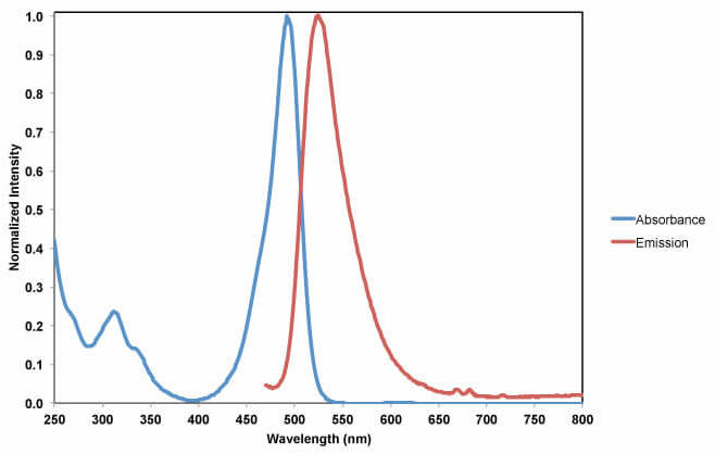
| Dye | Absmax | Emmax | ε(M-1cm-1) | MW (g/mole) | CF* |
|---|---|---|---|---|---|
| VRDye 490 | 491 | 515 | 73,000 | 1,011 | 0.11 |
*CF is a correction factor for the absorbance of the dye at Absmax to the absorbance of the protein at 280 nm.