Accurate, Replicable Western Blots with the Odyssey DLx

Odyssey® DLx
Imaging System

Get clearer answers with accurate, replicable Western blots and many other assays.

Get a Quote
Odyssey DLx

A Solution for Researching COVID-19

LI-COR considers it a great privilege to provide the products, tools, and services researchers need to arm themselves for the fight against SARS-CoV-2 and other viruses.

The Odyssey DLx Viral Solution Package is currently available for priority delivery and at a reduced price. Installation and training can be done virtually with a Solutions & Support Scientist.

Contact us
Sars-Cov-2 Sars-Cov-2 Sars-Cov-2

*Viral Solution Package Includes

Systems

  • Odyssey DLx Imaging System
  • Odyssey DLx Accessory Pack
  • Desktop Computer
  • Startup Reagents

Software

  • LI-COR Acquisition Software
  • Empiria Studio® Software for Western blot Statistical Analysis (10 Users) (1 User)
  • Lambda U® On-Demand Western Blot Education Portal (10 Learning Paths)

Services

  • Virtual Installation
  • Basic Virtual Training with an Expert
  • Advanced Virtual In-Cell Western and Viral Assay Training with an Expert
  • Unlimited Access to LI-COR Expertise

Contact us for additional information about the Odyssey DLx Viral Solution package.

*Offer may not be available in all regions.

plate, membrane, gel, slides

Do more with digital fluorescence

Get consistent, accurate digital images, without the hassles and unpredictability of film – so you can analyze your data right away and plan your next experiment.

in-cell western DLx

In-Cell Western Assay

target western Odyssey Dlx

NIR Fluorescent Western Blot

emsa gel shift Odyssey Dlx

EMSA / Gel Shift Assay

tissue section Odyssey DLx

Tissue Section

Image courtesy of C. Kearn,
University of Washington

Go where your experiments take you

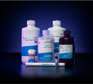
Western blots, cell-based assays, protein arrays, gel shift assays, tissue section imaging, and more are at your fingertips with near-infrared (NIR) fluorescence on the Odyssey DLx.

“We are finding that we can use the Odyssey CLx for just about everything protein-related we do in the lab.”

Dr. Jeremy Chambers, Florida International University

Image and analyze your samples quickly

Analyze your data right away – it’s already in digital format. One digital file contains all your image data. Record-keeping is easy and multiple exposures are a thing of the past.

To image and analyze many samples efficiently, use the Odyssey DLx to scan up to 9 mini-blots, 6 microplates, or 30 slides at the same time. The large scanning bed provides high throughput for many assays.


See the full story in a single image

See both strong and faint signals clearly in a single image acquired on the Odyssey DLx, with great sensitivity and no image saturation. Multiplex with two fluorescent colors to see even more data.

see more DLx

One digital image file contains all your data so you can see both strong and faint signals clearly in a single image, without image saturation, “blowout”, or sacrificing sensitivity. Increase the consistency and reliability of your results by capturing a single image with the Odyssey DLx, rather than comparing multiple exposures captured under variable conditions.

“The superior dynamic range and sensitivity allows me to confidently report my data.”

Lars Engstrom, Mirati Therapeutics, SelectScience® Review
Odyssey Dlx Western Blot Image Sequence 1 Odyssey Dlx Western Blot Image Sequence 2 Odyssey Dlx Western Blot Image Sequence 3 Odyssey Dlx Western Blot Image Sequence 4 Odyssey Dlx Western Blot Image Sequence 5 Odyssey Dlx Western Blot Image Sequence 6 Odyssey Dlx Western Blot Image Sequence 7 Odyssey Dlx Western Blot Image Sequence 8 Odyssey Dlx Western Blot Image Sequence 9 Odyssey Dlx Western Blot Image Sequence 10 Odyssey Dlx Western Blot Image Sequence 11 Odyssey Dlx Western Blot Image Sequence 12 Odyssey Dlx Western Blot Image Sequence 13 Odyssey Dlx Western Blot Image Sequence 14 Odyssey Dlx Western Blot Image Sequence 15 Odyssey Dlx Western Blot Image Sequence 16 Odyssey Dlx Western Blot Image Sequence 17 Odyssey Dlx Western Blot Image Sequence 18 Odyssey Dlx Western Blot Image Sequence 19

One image file contains the full range of your data, and changing image display settings never alters your raw data or signal intensities.

low background DLx

Get better sensitivity with low image background

Powerful, precise laser excitation and specialized optics enable the Odyssey DLx to provide high signal-to-noise ratios and outstanding image quality.

High signal-to-noise ratios give you more confidence in your data and can help you reliably detect subtle changes between samples. Low background fluorescence at NIR wavelengths means high sensitivity for your blots.

Visible region (Vy3) High autofluorescence Near-infrared region (700nm) Low autofluorescence

NIR fluorescence imaging gives you low membrane fluorescence and high sensitivity. Other imagers use LEDs or diffuse white light sources that provide weak, non-specific excitation light and limit sensitivity. Visible fluorescent dyes like Cy3 have poor sensitivity, because membrane autofluorescence is so strong. Blots show detection of phospho-ERK with NIR fluorescence (700 nm) and with ECL Plex reagents.

“The brilliant signal-to-noise ratio in combination with the ability to truly quantify the data is really outstanding.”

Geir Bjørkøy, University College of Sør-Trøndelag
sensitivity accuracy DLx

Enhance the accuracy of your data. The wide dynamic range of Odyssey imagers enables high-sensitivity detection without instrument saturation.

See your bands reliably with stable fluorescent signals

Fluorescent signals are stable and unaffected by timing, so you can compare band intensities with confidence. You can even conveniently re-image the same blot on the Odyssey DLx later and see the same results.

DLx stable signal

NIR fluorescent signals are stable for months, because no enzymes or substrates are used. Direct detection is performed using secondary antibodies labeled with near-infrared fluorescent dyes, such as IRDye® secondary antibodies. You’ll typically lose signal in less than an hour with ECL substrates and indirect enzymatic detection.

DLx direct indirect

Compare band intensities confidently with direct detection of stable fluorescent signals.

See multiple targets on the same blot

With multiplex fluorescence, you can detect two protein targets in each sample lane, with great sensitivity in both fluorescence channels.

Use secondary antibodies labeled with spectrally-distinct NIR fluorescent dyes to get more data from your blot. View, adjust, and analyze your results as a merged image, or as separate 700 nm and 800 nm channel images in pseudo-color or grayscale. Consistency and accuracy are improved because you can account for lane-to-lane variation in loading and transfer without stripping and re-probing.

“The infrared imaging system allows detection of multiple proteins on a single blot, without the need for stripping and re-probing.”

Bond et al (2008) Biol Proced Online. 10: 20–28.
DLx saturation

See your strong bands accurately

Get deeper data capacity with digital fluorescence and the unprecedented linear dynamic range of the Odyssey DLx, and never lose data because of image saturation.

Never lose your data because of image saturation. Digital fluorescent detection with the Odyssey DLx shows your strong signals as they really are. Film and CCD imagers only show you part of the picture, because data are lost when images become saturated. To see the whole picture, you need enough capacity (dynamic range) to consistently document your strongest bands.

“When saturated, film exposures can also hide sample-to-sample variations in high-abundance proteins.”

Janes (2015) Science Signaling 8(371): rs2
 
Film has a shallow data capacity and cannot accurately record strong signals. Once that capacity is reached, all additional signal (read: your data) is simply lost. Many CCD imagers are also limited by data capacity and image saturation.
data illustration

Acquisition and analysis software for the Odyssey DLx

Pair your Odyssey DLx Imager with Empiria Studio® Software for advanced acquisition and analysis using intuitive tools.

LI-COR Acquisition Software

LI-COR Acquisition Software

LI-COR Acquisition Software allows you to acquire, organize, and adjust images. A workflow-based system ensures no steps get missed, and you can acquire images using identical settings based on assay type. You can then export images for analysis in Empiria Studio or publication.

Empiria Studio

Empiria Studio Software

Empiria Studio enables you to perform in-depth analysis of images from a variety of assays. The intuitive tools in Empiria Studio minimize user-to-user variability, compute statistical values, and provide extensive analysis options for images acquired on your Odyssey DLx Imager.

Learn More about Empiria Studio

Odyssey DLx Imaging System

See the Odyssey DLx in action

Do more with consistent, robust digital imaging technology that has propelled the Odyssey imager family to over 20,000 peer-reviewed publications. See the benefits of stable NIR fluorescence and detect multiple targets in the same lane. Get the most accurate results with over 6 logs of linear dynamic range, so you can make more discoveries.

Need more information? Contact us.

* Remote demonstrations are available.

Looking to include chemiluminescent Western blotting?

With the Odyssey Duo, bundle an Odyssey DLx and an Odyssey XF together and save on the cost of purchasing each imager separately. Equip your lab with access to two near-infrared fluorescent Western blot imagers, a chemiluminescent Western blot imager, and an extended list of other applications.

Learn More