Revert™ 700 Total Protein Stain for Western Blot Normalization
Revert™ 700 Total Protein Stain Kits for Western Blot Normalization
Revert™ 700 Total Protein Stain and Wash Solution Kit
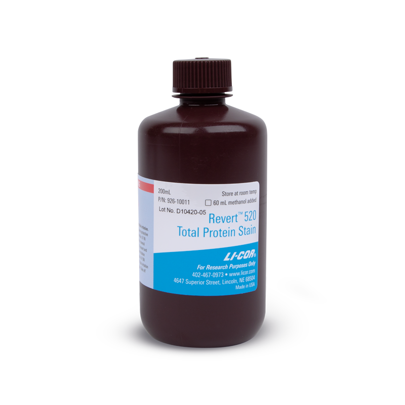
Revert™ 520 Total Protein Stain for Western Blot Normalization
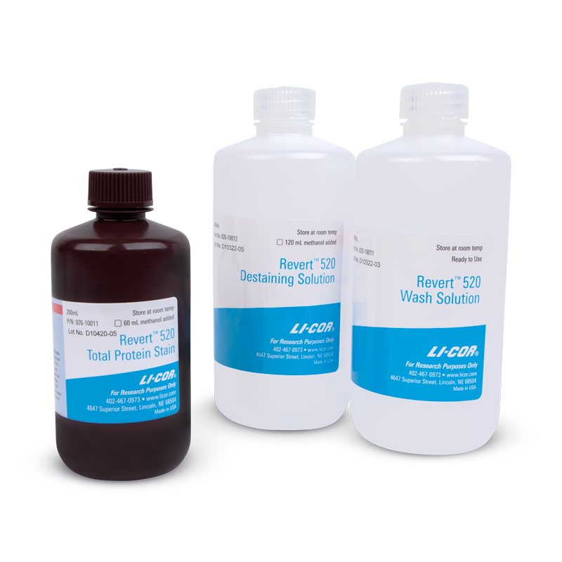
Revert™ 520 Total Protein Stain Kits for Western Blot Normalization
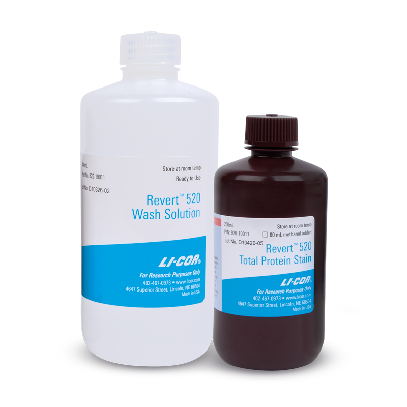
Revert™ 520 Total Protein Stain and Wash Solution Kit
Make Western blot normalization more accurate and reliable with Revert Total Protein Stain, a membrane-based (post-transfer) normalization strategy that stains all protein in your sample. No special reagents, equipment, or gels are required with Revert Total Protein Stains, so they are compatible with your Western blot protocol.
Total protein staining is considered the gold standard for Western blot normalization. Revert Total Protein Stains provide linear signal over a broad range of sample concentrations and are compatible with subsequent Western blot immunodetection methods.
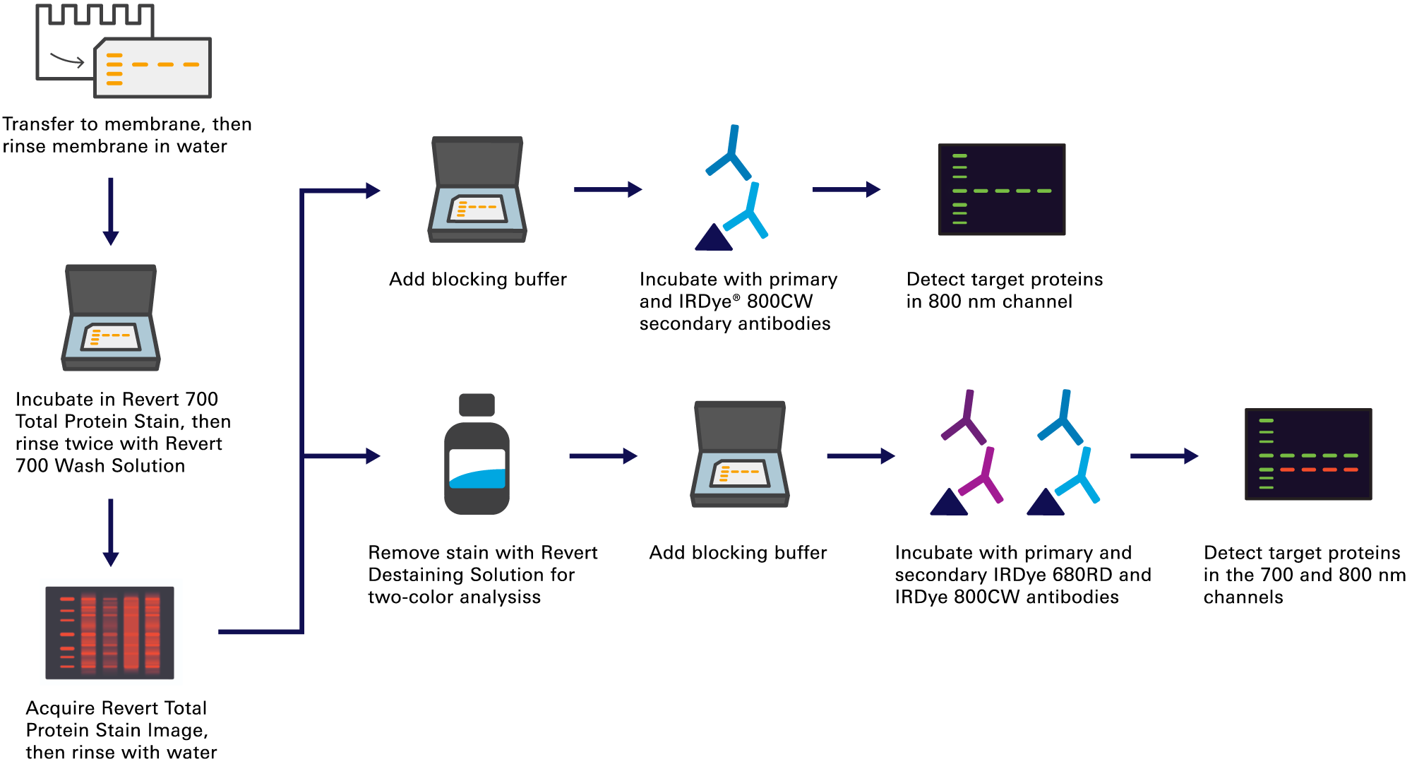
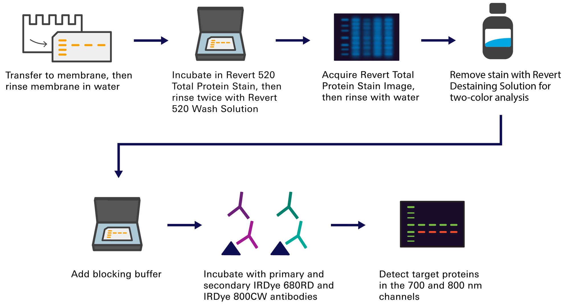
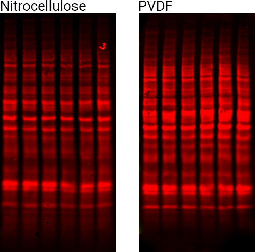
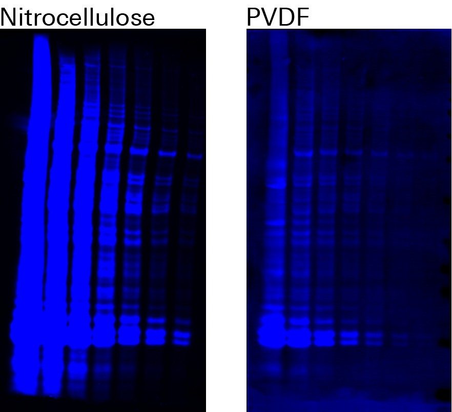
Revert Total Protein Stain works with your existing Western blot workflow, without needing any additional reagents or special gels. Staining takes less than ten minutes. (Figures 1 and 2).
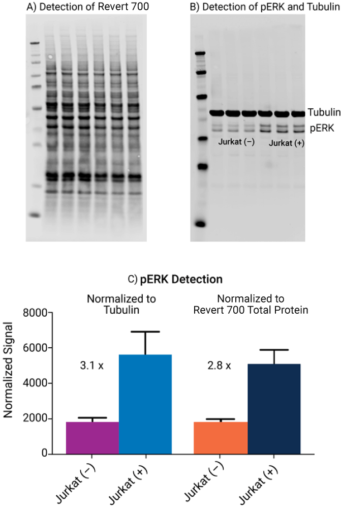
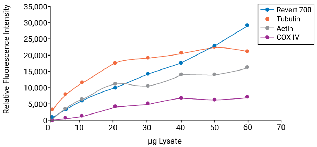
Use Revert stains to normalize accurately, by correcting for lane-to-lane variation in loading and transfer of target protein in your quantitative Western blots. Revert Total Protein Stains provide a linear response over a wider range of cell lysate concentration than some commonly-used housekeeping proteins (Figure 3).
Biological variation won’t affect total protein normalization with Revert. While validation of stable expression levels is highly recommended for housekeeping proteins, Revert Total Protein Stains are a consistent internal loading control that you don’t need to validate. (Figure 4).