To keep your targeted therapeutics moving through the development process, you need to be able to make decisions with confidence, and without hesitation or costly delays. Whether you’re characterizing a signaling pathway or validating a therapeutic candidate, confident decisions begin with exceptional data.

Get the answers you depend on to keep the development process moving forward. Odyssey® Imagers and Pearl® Trilogy Small Animal Imagers deliver superior data for robust, replicable results you can rely on.
IRDye® Reagents are optimized for use with Odyssey Imagers and the Pearl Trilogy Imagers to provide an additional layer of certainty.
Characterize your cell signaling pathways with confidence using quantitative, near-infrared (NIR) Western blots performed on an Odyssey Imager and quantitative, NIR In-Cell Western™ or On-Cell Western assays performed on an Odyssey M or Odyssey DLx. Gain a fuller understanding of the details of your ligands, targets, pathways, and cellular effects.

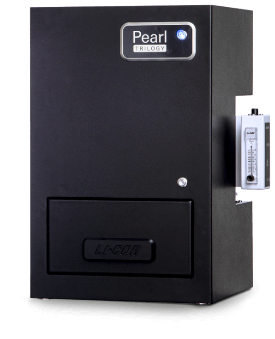
Assess your therapeutic agent’s performance in vitro and ex vivo using the Odyssey M or Odyssey DLx Imager, or in vivo and ex vivo using the Pearl Trilogy Small Animal Imager. Decide which of your candidates appears most promising and move to the next step of validation.

Understand more about your targets, pathways, and the effects your therapeutic has when you perform cell signaling assays on an Odyssey M or Odyssey DLx Imager.
Trust your quantitative Western blots, In-Cell Western assays, and On-Cell Western assays to give you the most reliable results when you use an Odyssey M or Odyssey DLx Imager and IRDye Reagents.
Use the IC50 assay with the Odyssey M or Odyssey DLx and IRDye Reagents to find the concentration of an inhibitor required to reduce a response by half. Get the data you need to continue through discovery and development.
PTK6 is abnormally expressed in breast cancers, and researchers have described kinase-dependent functions of PTK6 in driving tumor growth. As with any therapeutic approach, using small molecule inhibitors to target PTK6 activity requires validation of specificity and efficacy.
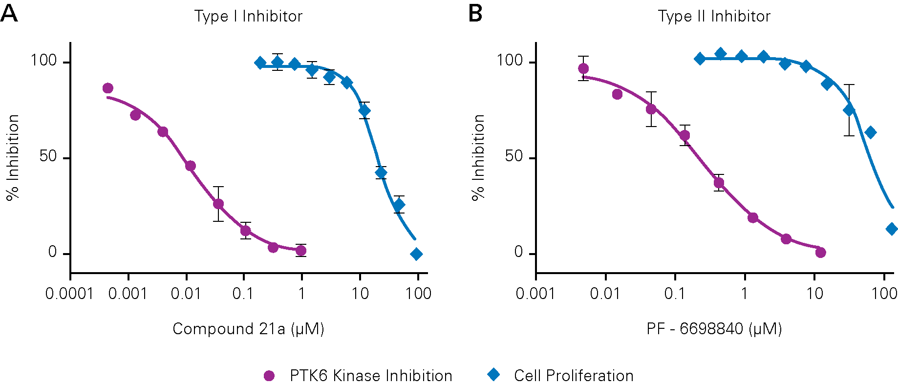
Inhibition of tumor cell growth by PTK6 inhibitors is independent of PTK6 kinase activity inhibition, providing evidence against using PTK6 kinase inhibition as a therapeutic strategy for breast cancer treatment. In-Cell Western Assays imaged on an Odyssey CLx Imager were used to assess cellular levels of PTK6 autophosphorylation normalized to cell number (measured by CellTag 700 Stain). Adapted from Qiu, L., et al.
From advanced analysis of vetted therapeutic leads to small animal, tissue, and organ imaging, we offer solutions to help you understand the key characteristics of your therapeutic before moving to the clinic.
The Odyssey M or Odyssey DLx Imager and the Pearl Trilogy Small Animal Imager provide an ideal combination to assess therapeutic delivery with NIR fluorescence and for complete analysis of your therapeutic agent’s performance.
The therapeutic agents tagged with IRDye Reagents deliver a clear image for detection and quantification. Validate your therapeutics and probes in vitro and ex vivo with the Odyssey M or Odyssey DLx, or in vivo and ex vivo with the Pearl Trilogy Small Animal Imager.
Learn how to maximize your targeted therapeutics research and make groundbreaking scientific advancements with NIR detection and innovative probe development.
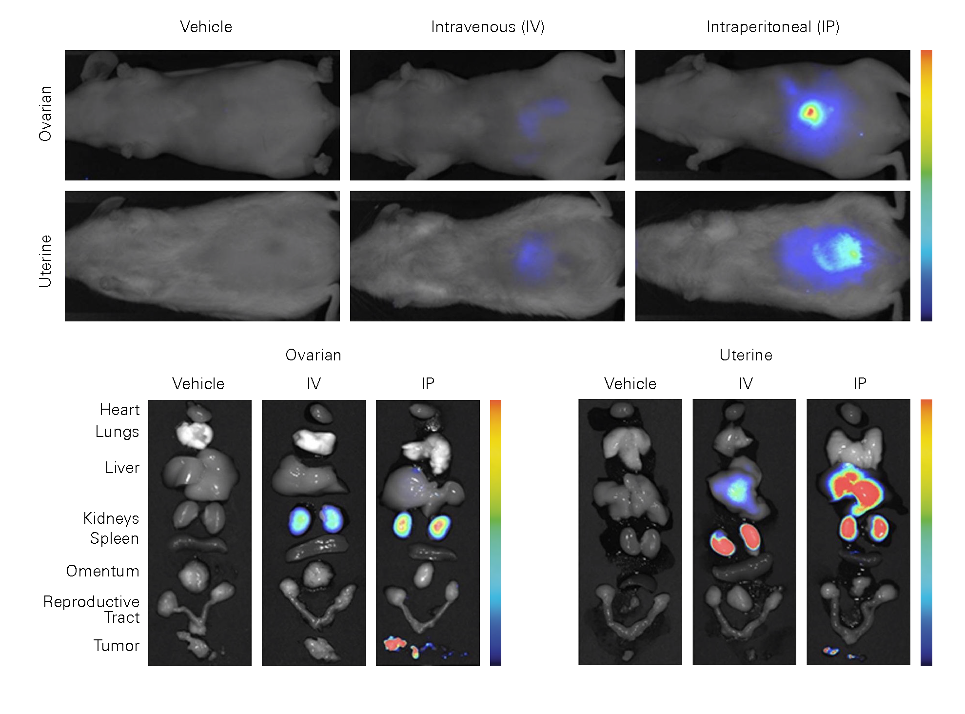
Ovarian and uterine cancers are often diagnosed at advanced stages, underscoring the need to identify therapeutic targets and means of delivering therapies to tumors. One promising therapeutic target is the AXL protein, which is involved in ovarian and uterine cancer metastasis. One potential treatment option is to silence AXL expression with siRNA, but a mechanism is needed to deliver the siRNA to the tumor.

Characterize and validate the specificity, binding affinity, and other parameters of your therapeutic agents with the In-Cell Western or On-Cell Western assay performed on the Odyssey M or Odyssey DLx Imager. Get the confidence you need in your agents before moving forward with small animal and tissue section imaging.

Capture a detailed representation of the in vivo molecular activity of your therapeutic agents in live animals. Assess uptake, clearance, and other key factors for development.

Use an Odyssey M, Odyssey DLx, or Pearl Trilogy Imager to quantify the presence of dye-tagged probes in excised tissue or organ samples to assess the biodistribution, clearance, and other parameters of your therapeutic leads.
From academic discovery to pre-clinical validation, LI-COR has the products, tools, and services to let you make critical decisions with confidence.
Get a Quote